Cerebrospinal fluid (CSF) is the clear, colorless liquid that surrounds the brain and spinal cord and is contained within a lining called the dura. The cerebrospinal fluid protects and cushions the brain and central nervous system. Among other functions, this fluid provides buoyancy to the brain, allowing it to float and weigh less, thus reducing the pressure at the base of the brain. A cerebrospinal fluid (CSF) leak occurs when there is a tear or hole in the dura that then allows this fluid to escape[1]. When leaks occur, the overall volume and pressure within the skull drops, and the cushioning and buoyancy effect is reduced, causing the brain to slump. In many cases, this leads to a condition known as intracranial hypotension and a vast range of symptoms.

The main symptom of a CSF leak is a headache that is worse when upright and improves when lying down horizontally. This is sometimes called a “positional” or “orthostatic” headache. However, not all positional headaches can be attributed to a CSF leak, and not all CSF leak headaches are positional. This is particularly the case in the chronic (vs acute) phase of CSF leaks, where the “positional” or “orthostatic” characteristic of symptoms may become more constant, lessen, or disappear entirely, including headache. Symptoms often worsen as the day goes on. Other leak symptoms can include, but are not limited to: nausea, vomiting, neck pain or stiffness, heaviness of head, pain between the shoulder blades, feeling of pressure within the head, changes in hearing (muffled or underwater sensation), tinnitus (ringing, buzzing, or pulsatile), feeling of liquid in the ears, sense of imbalance, sensitivity to light, sensitivity to sound, pain or numbness in the arms, changes in cognition (“brain fog,” memory loss, or loss of concentration), dizziness or vertigo, scalp sensitivity or tingling sensation within the scalp, visual changes (blurring, double vision, visual field defects), pain behind the eyes or when moving eyes, facial numbness or pain, sinus pressure, temporomandibular joint pain and stiffness, and subdural hematoma[2]. Cranial leak specific symptoms can vary even more and can include: fluid discharge from ears, nose (usually only one side) and to back of throat often reported as salty or metallic tasting, recurring or chronic meningitis, loss of sense of smell, change in hearing or ringing in the ears, and less frequently cognitive changes. Rare signs or complications of CSF leaks can include: quadriplegia, dementia (often mimicking Frontotemporal Lobe Dementia), Parkinsonism, other movement disorders, ataxia (unsteady gait), hypersomnolence, stupor, coma, stroke (hemorrhagic or ischemic), and even death.
CSF leaks are often very hard to locate, if ever. Approximately 50% of leaks cannot be found on imaging. Imaging and other tests used to attempt to find leaks are often read as “normal” even when there is a leak present. Other times, especially (but not always) in the case of chronic leaks, the positional symptoms either lessen or go away altogether, including the headache. Many who are leaking are not even aware that they are leaking. Leaks are often misdiagnosed as well[3]. Some of those common misdiagnoses are Postural Orthostatic Tachycardia Syndrome (POTS), migraines, sinus headaches, Meniere’s Disease, Chronic Fatigue Syndrome, Parkinson’s Disease (sometimes other neurodegenerative diseases), Fibromyalgia, Ehlers-Danlos Syndrome, Tarlov Cyst, Chiari Malformation, Cervical Spine Disease, cervicogenic headache, tension headache, and Sinusitis. To make diagnosis even more complex and elusive, CSF leaks can and do often occur along with any of these disorders and perhaps several simultaneously. A leak can cause an acquired Chiari malformation or coexist and complicate an existing congenital Chiari malformation[4]. Some patients have had unnecessary decompression surgeries when the underlying, sole cause was a leak all along.
Leaks can be caused by:
- Medical procedures (also called iatrogenic leaks) for various diagnostic or therapeutic reasons such as lumbar punctures to collect fluid for analysis if meningitis is suspected, lumbar puncture for injection of contrast (myelography), spinal anesthesia, epidural injections, epidural steroid injections, prior skull base or spinal surgery, CSF shunt over-drainage, prior sinonasal surgery, and chiropractic or other spinal manipulations.
- Traumatic injuries such as brachial plexus injuries falls, sports injuries, motor vehicle accidents, roller coaster rides, and other whiplash injuries.
- Spontaneous leaks that occur with minimal or no clear cause. Sometimes spontaneous leaks may be associated with some sort of spinal pathologies such as calcified disk material or bone spurs. These leaks are usually ventral (or in front of the spinal cord).
There is growing evidence suggesting that a significant number of spontaneous CSF leaks occur as the result of a preexisting weakness in the dura[5]. Heritable Disorders of Connective Tissues (HDCT’s) such as Marfan Syndrome, Ehlers-Danlos Syndrome (both classical and hypermobility type), autosomal dominant Polycystic Kidney Disease, and other HDCT’s predispose patients to CSF leaks. One leak expert estimates that “slightly less than 100% of patients with spontaneous CSF leaks have an underlying connective tissue disorder.”[6] The dura is made out of connective tissue and patients with HDCT’s have thinner dura mater, that is more susceptible to tears and leaks. HDTC patients are more prone to spinal conditions such as perineural cysts, meningeal diverticula, and other HDCT defects such as aneurysms and dilatations. Oftentimes, a CSF leak is the first sign of an underlying HDCT.
Lumbar punctures (LP’s or spinal taps) should be avoided in patients with Chiari Malformation and/or in patients with HDCT’s[7]. There is a risk of causing a herniation of the cerebellar tonsils or making an existing herniation worse from the pull-down mechanism involved in lumbar punctures. Unfortunately, lumbar punctures are not always avoidable and sometimes very necessary, especially in cases to rule out life-threatening viral or bacterial conditions such as meningitis, subarachnoid hemorrhage, encephalitis, or syphilis. In these cases, measures can be taken to minimize LP risks such as using a certain needle type and size, limit the number of cc’s collected (by spontaneous drip ONLY), and, of course, always done under fluoroscopy by a competent physician[8]. Additionally, it is important to be aware that patients with HDCT’s are at greater risk for the dura to fail to heal following an LP. Patient’s should be aware of post-dural puncture headache (PDPH) symptoms and speak with their physicians if they suspect a leak following an LP.
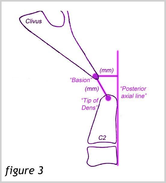
The procedures and tests used to diagnose leaks will vary between patients and certain criteria are used to diagnose leaks[9]. Some of these tests and procedures might be: endoscopic exam and fluid collection and Beta-2-Trasnferrin testing (cranial leaks), Cisternography (including radioisotope cisternogram), Magnetic Resonance Imaging (MRI) including Magnetic Resonance (MR) myelography, Dynamic CT myelography, Digital Subtraction myelography, and Intrathecal saline infusion-enhanced myelography, a lumbar puncture to collect and test fluid and measure opening pressure. This imaging often includes both brain and spinal imaging. The normal opening pressure is not uncommon and does not rule out a leak. High pressure can also occur while leaking. Pre-existing intracranial hypertension can be related to the development of spontaneous spinal CSF leaks. Some reports suggest that spontaneous cerebrospinal fluid (CSF) leaks are strongly associated with idiopathic intracranial hypertension (IIH). There are 5 main findings on imaging that doctors look for, however, the absence of these findings does not rule out a CSF leak. The mnemonic SEEPS is used for most of these findings: subdural fluid collection, enhancement of pachymeninges, engorgement of venous structures, pituitary hyperemia, and sagging of the brain[10]. Other imaging findings that might be seen are small ventricles, cisterns might have less fluid, optic chasm might flatten over pituitary, pituitary might enlarge, empty sella, fluid in front of the pons, or pons might become flatter than normal. Repeat imaging is often necessary.
Treatment of leaks can either be medical or surgical. Conservative treatment is often recommended, if possible. This can include bed rest and avoidance of coughing, sneezing, straining, bending, twisting, and lifting, increased fluid intake and caffeine, the use of an abdominal binder, and sometimes steroids are recommended. Others may have a lumbar drain placed in the low back to decrease the pressure of the CSF around the area of the leak in an attempt to allow this area to heal. Some patients may need an epidural blood patch. Where a blood patch is not successful, a fibrin glue patch may be tried. About 30-40% of leaks occur at multiple sites, especially in those with an HDCT. Multisite patches may be required. Higher volumes of blood may be needed in order to reach where it needs to go, and/or the position of the needle may need to vary from the standard placement (transparietal or lateral placement)[11]. It is not uncommon for several patches to be tried. Many doctors make the mistake that if an EBP fails, there was no leak as well. Sometimes when more conservative and less invasive treatments have failed, neurosurgery may be necessary. Surgical repairs vary and are tailored according to the type and location of the leak. Sometimes in a select set of patients, other procedures have been used including epidural saline infusions through an implanted epidural catheter or lumbar dural reduction surgery. A condition known as rebound intracranial hypertension (RHP) may occur following any of these treatments[12]. Usually, but not always, there is a different pattern to the headache where one feels worse when horizontal and better when upright. Sometimes, acetazolamide (Diamox) or a similar medication is prescribed to help treat RHP.
Leaks are poorly recognized, poorly understood, under-researched, understudied, often misdiagnosed, can complicate existing conditions, are difficult to find, mimic many other disorders (including Chiari Malformation), and can be comprised of a vast array of symptoms. Most doctors are familiar with the symptoms of a leak in the acute phase. Very few doctors are familiar with how long-term, chronic CSF leaks “present” in regard to headaches and other leak symptoms and often miss the more subtle symptoms of chronic leaks. Like Chiari and other related disorders, no two patients with CSF leaks have the same symptoms and often experience misdiagnosis, delayed diagnosis, are disbelieved concerning their symptoms of the severity thereof, and all too often dismissed to suffer excruciating pain, decline, and debility. Educating yourself as much as possible about CSF leaks will help guide and empower you and those around you who may have existing, suspected or potential future complications that may arise due to CSF leaks.
CSF Leak Symptoms List: https://dev.chiaribridges.org/glossary/symptoms-of-intracranial-hypotension/
[wpedon id=”4396″ align=”center”]
References:
1 “CSF Leak: A Curable Cause of Headache.” A Non-Profit Hospital in Los Angeles, <www.cedars-sinai.edu/Patients/Programs-and-Services/Neurosurgery/Centers-and-Programs/Cerebrospinal-Fluid-Leak/CSF-Leak-A-Curable-Cause-of-Headache.aspx>.
2 “Cerebrospinal Fluid (CSF) Leak.” A Non-Profit Hospital in Los Angeles, <www.cedars-sinai.edu/Patients/Health-Conditions/Cerebrospinal-Fluid-CSF-Leak.aspx>.
3 Schievink, Wouter l. “Misdiagnosis of Spontaneous Intracranial Hypotension.” Archives of Neurology, American Medical Association, 1 Dec. 2003, <www.jamanetwork.com/journals/jamaneurology/fullarticle/785098>.
4 Haider, Ali S., et al. “Spontaneous Intracranial Hypotension Presenting as a ‘Pseudo-Chiari 1.”Cureus, Cureus, 16 Feb. 2017, <www.ncbi.nlm.nih.gov/pmc/articles/PMC5354398/>.
5 Reinstein, Eyal, et al. “Connective Tissue Spectrum Abnormalities Associated with Spontaneous Cerebrospinal Fluid Leaks: a Prospective Study.” European Journal of Human Genetics, Nature Publishing Group, Apr. 2013, <www.ncbi.nlm.nih.gov/pmc/articles/PMC3598315/>.
6 Schievink, Wouter l. “Q & A With Dr. Schievink.” Spinal CSF Leak Foundation, 14 July 2014, <www.spinalcsfleak.org/q-a-with-dr-schievink/>.
7 Erbay, Sami H., et al. “Is Lumbar Puncture Contraindicated in Patients with Chiari I Malformation?” American Journal of Neuroradiology, American Journal of Neuroradiology, 1 Apr. 2005, <www.ajnr.org/content/26/4/985>.
8 Matas, Sandro Luiz de Andrade. “Why Should We Use Atraumatic Needles in Lumbar Puncture?” Arquivos De Neuro-Psiquiatria, <www.scielo.br/pdf/anp/v71n9B/0004-282X-anp-71-09b-681.pdf>.
9 Schievink, W. l., et al. “Diagnostic Criteria for Spontaneous Spinal CSF Leaks and Intracranial Hypotension.” American Journal of Neuroradiology, American Journal of Neuroradiology, 1 May 2008, <www.ajnr.org/content/29/5/853>.
10 Schievink, Wouter l. “Spontaneous Spinal Cerebrospinal Fluid Leaks and Intracranial Hypotension.” JAMA, American Medical Association, 17 May 2006, <www.jamanetwork.com/journals/jama/fullarticle/202849>.
11 Griauzde, J., et al. “Large-Volume Blood Patch to Multiple Sites in the Epidural Space through a Single-Catheter Access Site for Treatment of Spontaneous Intracranial Hypotension.”American Journal of Neuroradiology, American Journal of Neuroradiology, 30 Apr. 2014, <www.ajnr.org/content/early/2014/04/30/ajnr.A3945>.
12 Kranz, P. G., et al. “Rebound Intracranial Hypertension: A Complication of Epidural Blood Patching for Intracranial Hypotension.” American Journal of Neuroradiology, American Journal of Neuroradiology, 1 June 2014, <www.ajnr.org/content/35/6/1237>.
 Pain from a Chiari headache can be brought on from the simplest of things – a sneeze, a cough, laughter, or bearing down when going to the bathroom. We never know when the headache is going to strike, how long it will last, or when it will end. We are unable to describe the intensity of the pain to others, and when asked to rate our pain on a scale of 1 – 10, we want to scream, “14!” The radiating, crushing pain of the headaches robs us of our ability to function for days on end. And depending on the extent of damage the Chiari has caused you may also have the burning, stabbing, and shooting involved with neuropathic pain and neuralgia. The frosting on the pain cake, perhaps? With the many co-morbid disorders that go hand in hand with Chiari, such as Ehlers-Danlos Syndrome, Intracranial Hypertension, Hydrocephalus, and Tethered Cord Syndrome, is it any wonder we cry out, “Make it stop!”?
 By a wide margin, the hardest part of our fight is dealing with doctors. One would think it would be the never-ending pain, but when your doctors do not believe you, ridicule you, or outright verbally abuse you, it not only adds insult to injury, but it probably illustrates reasons that your doctor(s) just might be the ones that need counseling. As patients, we are paying for them to help us with our medical problems; what we get instead is usually a referral to a therapist because of their ineptitude. The absurd part of this circle of insanity is that when we make ourselves more knowledgeable about our condition(s), because we have no other choice, a doctor that understand the Hippocratic Oath would respond by making themselves more knowledgeable as well, so they can help their patients. Yet, what is far too common are doctors that do not want to know the results of studies, who are complacent with their fifteen minutes of Chiari education, and think if they talk a good game, patients can be manipulated into thinking maybe it’s all in their minds.
 In a distant second, would have to be the heart-wrenching feeling we get when our loved ones put more stock in the opinions of our morally bankrupt doctors, even once we have shared study after study and article after article showing you that our doctors are wrong. What we go through, feeling like our bodies have betrayed us, knowing that our doctors have betrayed us (even if it is because they do not know any better), we need you in our corner. This is the fight of our lives, for our lives, and we should never have to do it alone! The energetic, feisty, loving person that you have loved so much over the years is still inside of us! When your brain falls into your
  (apparently, they do not know the importance of the Central Nervous System either). Although benefits can still be fought for and awarded, the fight becomes far more difficult than it needs to be. Chiarians often must fight for years, filing appeal after appeal, to prove that their reality is the reality.
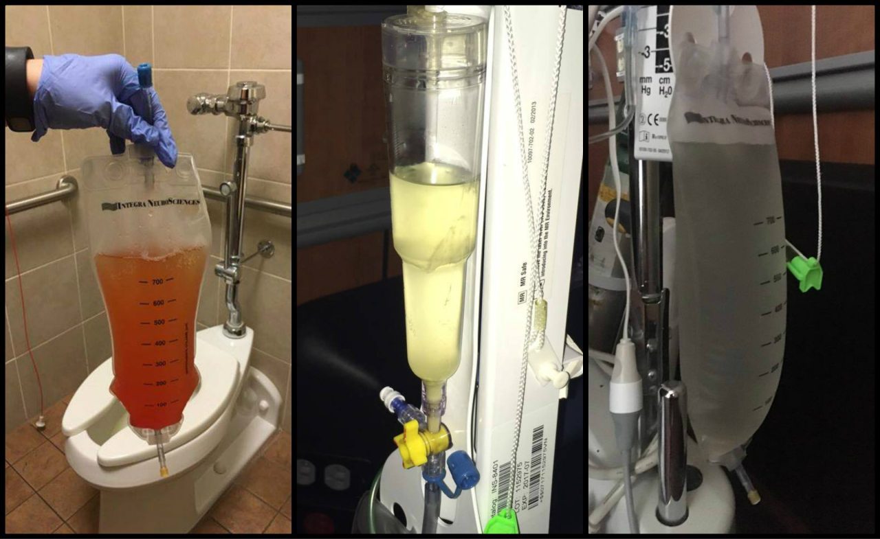
![The Christopher Ellington Story – A Chiari Warrior’s Journey [Updated]](https://dev.chiaribridges.org/wp-content/uploads/2017/05/hosp1.jpg)

 One of the biggest hurdles a Chiari patient may face is that of simply being diagnosed. Some studies cite an average of 5 years between the onset of symptoms significant enough for a patient to seek medical care and the patient receiving an accurate diagnosis of Chiari Malformation. Sadly, however, online support groups and message boards are peppered with the stories of patients who went undiagnosed, or more often, misdiagnosed, for decades. Patients are frequently misdiagnosed with conditions such as Fibromyalgia, Multiple Sclerosis, Chronic Fatigue Syndrome, Chronic Migraine, and various autoimmune disorders. Even more disturbing is the fact that in a study by Dr. Thomas Milhorat of over 300 patients diagnosed with Chiari, 59% had been diagnosed with a psychosomatic illness.
 Diagnosis of Chiari Malformation is based upon the presence of Chiari symptoms, such as an occipital headache that is brought about or worsened by Valsalva maneuvers, neurological symptoms such as poor balance, numbness or tingling in the arms, weakness in the legs, etc. combined with the “gold standard” of imaging studies for Chiari, an MRI of the brain.
 becoming apparent that Chiari is not quite as rare as it was once thought to be. When a primary care physician is stumped by a patient’s complaints of headaches and neurological symptoms, it is only natural to refer that patient to a neurologist for evaluation. But even many neurologists are grossly uninformed about Chiari. Many patients, even with an MRI that shows a herniation, are told that their many varied symptoms can’t possibly be due to the herniation of their brain, even with numerous studies available which show otherwise. Some patients have even been told by neurologists and even neurosurgeons that “Chiari doesn’t cause headaches [pain].” In fact, most symptomatic patients experience severe headaches, nerve pain, and pain from related disorders such as Syringomyelia.
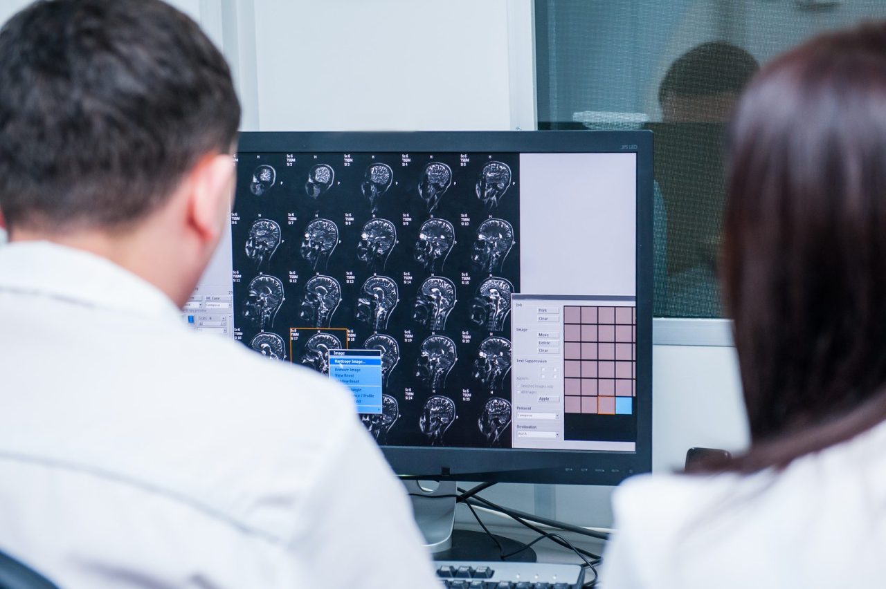
 Once diagnosed, you will usually be referred to a specialist (not a Chiari Specialist, but an everyday, run-of-the-mill neurologist or neurosurgeon). They tend to come in one of two types: Either they are very passive and just want to wait and see how bad it gets, or they are very pro-surgery and while they will still usually give you a 50% chance of helping your symptoms, they will tell you how decompression surgery really is your best option. Both are problematic.