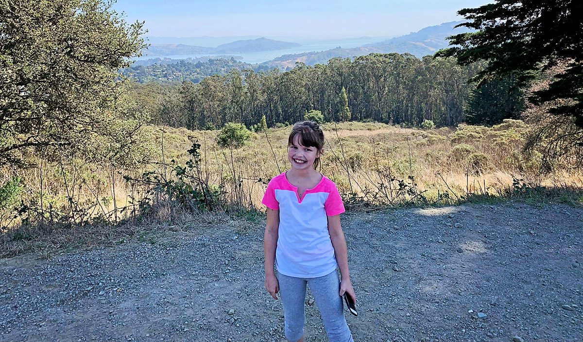
Colton had just finished his Chiari Decompression. He was headache free and doing great! His neurosurgeon came to me and said, “This is a genetic disorder and Emmalyn should be checked.” Two weeks later I took Emmalyn into the Chicago area to be sedated for her first MRI of the brain and spine. Three hours later I met Emmalyn in recovery where she came out of anesthesia smiling. Emmalyn was three years old and asymptomatic, not even a headache. Colton’s neurosurgeon called me the next day and let me know that she was only 5mm herniated, but in the spinal MRI, she had two separate syrinxes. A smaller one in her cervical spine and a very large one in her thoracic spine. Her opinion was to decompress Emmalyn right away as she was very concerned about the two syringes and explained, “With the decompression, it should allow the syringes to dissipate.” So, we scheduled her first decompression for two-weeks later, on July 31st, 2012. Emmalyn went through her first decompression like a champ and was released from hospital after three days. This is when Emmalyn’s headaches started. It seemed like once a day she would be getting a headache. Two weeks into recovery Emmalyn was sitting in a chair and said that she felt sick to her stomach, so she ran into the bathroom, I followed and as she was heaving her head went deeper into the toilet. I picked her up and her eyes darted to the right and she couldn’t talk to me. I immediately called 911. When the paramedics arrived, they swept her from my arms and rushed her to the ambulance. They kept her in front of our house in the ambulance for about ten minutes before they came and told me she was having a seizure; they didn’t want to jostle her head, so they called the helicopter. We were told to meet the helicopter at the hospital, which is forty-five minutes away. My husband and I jumped in our van and I don’t think I have ever seen my husband drive so fast; we beat the helicopter there. When the helicopter landed, we were in the ER to greet her when she came off the elevator, and when she did, she was covered in blood and screaming. I looked to the helicopter nurse and she said, “Two minutes before landing she came to and she wanted her mom and pulled out her IV.” She had a forty-five-minute seizure. We are in “small-town,” USA, so the ER did a quick MRI, bloodwork, but no EEG and at the time I didn’t know she should have had one. So, they said she was fine and released her from the hospital. Of course, after speaking with her neurosurgeon the next day she had us in the car to make the five-hour drive for her to be admitted. After three days of tests, they concluded that she had chemical meningitis and put her on medication and anti-seizure meds for six months. Let me tell you that was the scariest time of my life, but Emmalyn took it like a champ. In three months, we had repeat scans, her decompression site looked good, but her syringes didn’t change in size, so we chose to wait and see.

In 2013, Emmalyn developed leg pain and started having incontinence. She potty trained early and never had problems. After going through more imaging and urodynamics they said that she didn’t look like she was tethered but they were sure that she was. They called it Occult Tethered Cord. So, Emmalyn underwent a tethered cord release on September 27, 2013. She only spent one night in the hospital and was up and doing all well, so they let her go home. After the release, her incontinence subsided but the leg pain continued.

Over the next two years, we monitored Emmalyn and her headaches continued. In 2014, we did our next repeat MRI and it showed that Emmalyn’s syrinx in her thoracic spine had gotten a little longer in length. It concerned her then neurosurgeon and she made the decision that we should do another decompression in hopes to reduce the size of Emmalyn’s syrinx. On July 1st, 2014 Emmalyn went for her second decompression. Once again, she came through everything like a champ. One thing about Emmalyn she is a fighter!

For the next year Emmalyn struggled with more headaches and leg pain, so more imaging was done. She had developed scar tissue that was blocking her CSF flow once again. After the imaging, the decision was made that in November, she would undergo yet another decompression to clean up the scar tissue so that flow could be reestablished. Emmalyn underwent her third decompression on November 2nd, 2015. She sailed through surgery and the surgeon came out to talk with my dad and me. She explained that Emmalyn’s future could be complicated as she was developing a lot of scar tissue and that would make things more complex because it could continue to block the flow. The pain after these surgeries is something, they don’t prepare you for and after this surgery, it was worse than the last two. She came out swinging and hated everything. They got her pain under control, but it seemed like more pain medication was needed this time to keep it that way. This time, she spent four days in the hospital before returning home.

After the surgery, her pain got a little better. Then on December 29th, our world got turned upside down. We were at my niece’s house in Wisconsin and my mom had run her hand down the back of Emmalyn’s head and she yelled for me to come over there. Emmalyn had a large lump on the back of her head that was squishy. We rushed to the ER in Madison and they took her straight to MRI and that is when we found out Emmalyn had developed her first pseudomeningocele. It was very large. They only let us leave with her when I promised to get an appointment with her neurosurgeon within the next week. On a good note, Emmalyn won her first American Girl Doll Grace while in the ER. She was so excited. It brought a smile to her face in between the bad headaches she was having. At this point in life, Emmalyn had lived with two years of headaches and had become known as the girl with a smile. She always smiled through her pain.

On January 5th, 2016 we took the five-hour drive back to her neurosurgeon. She took one look at the back of her head, and the imaging and she said we would be going back into surgery the next day to repair. Emmalyn was an add-on to their schedule, so she wouldn’t be going to surgery until noon. Keeping a five-year-old with no food or drink after midnight to noon is quite a task, but when the prior surgeries took longer than they thought, going until 5 pm was more than a task. She was mad, tired, hungry and beyond over it. They came to get her and even with all she had been through, she smiled and said, “love you all, see you when I wake up.” She was always so easy going into the operating room. After three hours, her neurosurgeon came out and said there was a pin-sized-hole in the dura. She showed me a picture, and she repaired it with a patch. Once again in true Emmalyn fashion, she woke up swinging, mad and in pain. By this point, they had her pain regimen down to a T, which is SO important. Her recovery went well, and she was out in three days and sent home.

Once again, her headaches and leg pain continued, but she was able to be upright again. In the middle of February, the back of her head became squishy again and her headaches started to get worse when upright. I called her neurosurgeon, and as always, more imaging was ordered, and as I suspected, her pseudomeningocele was back again. She delayed surgery as she wanted to do some research. So, Emmalyn laid down for most of a month. On March 31, we returned to the hospital for her sixth surgery. She went in and did the repair and placed an EVD drain, to check the pressure and drain CSF fluid. The drain was placed for a week, and it was the happiest and pain-free I had seen Emmalyn in over two years. Her pressures were all above 28, which meant high pressure. She made the decision that a VP shunt needed to be placed. Emmalyn went in for her seventh surgery on April 6th. Her Neurosurgeon came out and talked to me and my parents, she let me know that if this patch was to blow and another pseudomeningocele was to appear that at this point she wouldn’t know what to do next, that Emmalyn was too complex for her. I wanted to cry in fear for the first time. My dad said then what, whatever it was we would make it happen. Thank God for my dad, my rock. She said that she would refer us to a specialist she knew of in NYC. I prayed right there this night for god to take care of my little girl and heal her. After she woke up, the pain wasn’t as bad. Shunt placement seemed to be much easier than decompression. She stayed for two more days and was released to return home.

Emmalyn’s headaches continued, and one month later her pseudomeningocele reared its ugly head. I called her neurosurgeon and she did imaging and this leak was bigger than the other two. It consumed the whole back of her head. She made the referral to the specialist in New York City, NY.

After sending all her imaging, op notes, doctor’s notes, and everything else. We consulted with the specialist’s office. An appointment was made to go to NYC and see him on June 10th, 2016. My sister, Emmalyn and I boarded the plane for our first trip ever to NYC. We met with the specialist, who went over all of Emmalyn’s imaging, talked with all of us, and checked Emmalyn over. He let us know that Emmalyn was a very complex case, that her first neurosurgeon had removed too much bone and most likely cause her to have Craniocervical Instability (CCI). He also said that he suspected that she had a connective tissue disorder called Ehlers-Danlos Syndrome (EDS), and that was why healing had been hard and why the dural patch was not holding. His surgical plan was to go in and once again patch the leak in the dura, clean up the scar tissue, place a titanium plate to hold where her brain was slumping, place an EVD drain again for a week to make sure the VP shunt was needed, and because of the EDS aspect to be closed by a plastic surgeon with muscle flaps to make sure it would hold. Before he would schedule surgery, he wanted to speak with her former neurosurgeon in depth to know what he was in for when he cut open the back of her head. He said that we could fly home and he would be in touch on when surgery would be scheduled.

Emmalyn struggled for a whole summer with severe headaches as we waited for surgery to be scheduled. Finally, at the beginning of September, we received the call that we would be returning to NYC on Sept 15, 2016, for pre-op and surgery on the 19th. Dreams come true and prayers answered. We would have to be in the city for two weeks, as she would be in the hospital for a little over one week. We returned to NYC and everything went as planned. She and I walked to the OR. She had a smile on her face, and she said, “I love you, mommy,” as she went under. As always Emmalyn woke up swinging and in so much pain. We were at a new hospital with nurses and staff that didn’t know her pain regimen, so momma bear kicked into action. At first, they wouldn’t listen when I told them that the only thing that worked for her for the first twenty-four hours after surgery was morphine, and then she could be switched to Oxycodone. Once I was finally heard Emmalyn’s pain was taken care of. Within forty-eight hours Emmalyn was up and walking with her drain and feeling great. Her pressures were still above 28 the whole week so the decision was made to put the VP shunt back in place with the valve set at 1.5. She returned to the OR on September 26th for the shunt and was discharged from the hospital the next day. The next sixty-eight days were the best days of my daughter’s life; she was finally pain-free!

On the 4th of December 2016, Emmalyn’s leg pain returned, and two days later her headaches returned when upright. I emailed her new neurosurgeon immediately. He ordered stat MRIs of the brain and complete spine. I received a copy of the MRIs on disc and looked at them when I got home, and my worst fears came true – the pseudomeningocele had returned. I emailed her neurosurgeon a few images and he called me right away. He explained that he believed the leak was back and he wanted us on a plane ASAP. We flew back to NYC in two days and my parents followed three days later. When we arrived, he admitted her immediately through the ER. He explained surgery would take place on December 14, 2016, for another pseudomeningocele repair. On the 13th of December, he came into her room and switched gears, he believed that her shunt was malfunctioning and there was not a leak. I told him that her symptoms were the same as every leak before and he said he was very sure it wasn’t; so, on the 14th they would do the surgery to check her shunt. He took her into surgery on the 14th and come out and said that there was nothing wrong with the shunt, but he dialed it to 0.5 to help with the CSF and we could return home after post-op.

We returned home in time for Christmas and Emmalyn’s headaches continued to be horrible. I kept in constant contact with her new neurosurgeon about it and he kept saying let’s wait and see. We ended up having some insurance troubles as his office no longer excepted our insurance, so we had to take out an additional private policy for Emmalyn to be able to continue to see him. After all that was solved, we returned to NYC at the beginning of March for an ICP bolt test. Emmalyn returned to the OR on March 2nd, 2017 to have an ICP bolt placed to once again check her pressures as he believed her current VP shunt wasn’t aggressive enough. Her pressures were still a little high but not like before. She was in the hospital for forty-eight hours with the bolt and then it was removed at her bedside. She screamed through the removal. He sat down with us at post-op and talked to us in-depth that he still believed that there was not a leak and that he wanted to place a new VP shunt, with no valve, but instead she would have an anti-siphoning device behind her ear to slow it down when she was upright. He believed this was the best course of action for Emmalyn. He is the specialist, so of course, why would I question it. So, we scheduled her next surgery for March 22nd.

We returned to NYC at the end of the month and she went back into her first surgery of two on March 22, 2017. In the first surgery, he placed an EVD drain to drain CSF and to check pressures again. Her pressures were normal but on the higher size so he proceeded to surgery number two on March 29th. He removed the EVD drain and placed the new no valve shunt and the anti-siphoning device that could be changed when needed. It was a very painful surgery for Emmalyn as the placement of the anti-siphoning device was behind her ear in a very tender spot, and he also let me know she was the first child he had ever placed this in. She had recovered and returned home. Her headaches continued.

We had to come back in May for imaging as no one in our area would do MRI’s on her now due to liability issues with the shunt. The pseudomeningocele was still present and her headaches were still strong. He made the decision to give it until July and if things hadn’t improved, he would open the back of her head and check for the leak.

We returned in July and nothing had improved. We sat down to discuss the next surgery and he switched gears again. He said after a discussion with his colleagues, he still didn’t think there was a leak. He then let me know he thought adding a lumbar shunt would help the situation to take care of the pain and the pseudomeningocele. Once again over my better judgment, he is the specialist, so we scheduled surgery, and her lumbar shunt was placed and set at 1.5. It was an easier surgery for her, so her hospital stay wasn’t as long. The next few days at the hotel were horrible her head pain was so she couldn’t even walk or be upright in bed without a debilitating headache. After speaking with her neurosurgeon, he said take her to the ER and have it dialed up to 2.0. Upon arrival at the ER, they checked her shunt and after her previous MRI, the resident dialed it to 0.5 rather than 1.5 (so it was virtually wide open). The current resident dialed it to 2.0 in front of me. Things improved. Recovery from this developed a whole new symptom – stomach pain. Now she had daily headaches, leg pain, and stomach pain, but through it all remained smiling as much as she could. He wanted her back in a month, for new imaging, to see how things were doing.

In a month nothing improved, she just continued to get worse. So, in August we returned for imaging and an appointment. The imaging showed that the pseudomeningocele had almost gone away. He was so happy. I was too, but her symptoms had not gone away at all and now the added stomach pain was causing even more suffering. So, he said we should return again the next month, and they would externalize the shunts to see if the stomach pain would go away and if it did, we would convert the VP to a VA to get the tubing out of her stomach.

We returned in September. He externalized her VP shunt but not the LP. Her stomach pain didn’t improve so he said it wasn’t the shunts, so he took her back into the OR to place her shunts back in place. Although I explained her headache pain was not better and I still believed she had a leak from her dura. He said he was positive there wasn’t a leak.

We returned to Illinois. Her symptoms continued to be debilitating. I emailed him at least five times a week for answers and he quit answering. So, I called his office and they would set up phone call appointments that he never kept. All attempts to contact him were ignored for three long months, while our eight-year-old Emmalyn suffered. Until the day I emailed his college about the problems she was having, asking, begging for anyone to give us a second opinion, and what do you know he called me ten minutes later. He promised he would come up with a plan, and said he thought she needed pain management as everything surgical was stable. After a week of hearing nothing, I emailed him one last time. Asking him to open the back of her head and check for a leak, if there wasn’t one, I would concede to pain management. His office called the next day to set up surgery.

We returned to NYC on December 10th for surgery on the 11th. He told me surgery should be less than two hours as he didn’t believe he would find anything. He came out to the waiting room five hours later. He took me outside the waiting room and explain to me that when he opened the back of her head, he found many holes in her dura that were causing a leak. He explained that he went further up on her head and harvested her own tissue and sealed the dura again. He told me that he believed this would secure the dura and we would never have a leak again. He said that the plastic surgeon was closing her up, and I should see her in about an hour. He assured us that he would be up the next day to talk further. What a punch in the gut. Emmalyn woke from surgery in so much pain, but the wonderful PICU team that knew her so well jumped into action. A lot of these nurses have at this point gone from just nurses to being like family to us. Our stay after this surgery was rough, she was hospitalized for ten days and couldn’t be upright longer than forty-five minutes without morphine. Her leaks were sealed, but with two shunts over-draining, her low-pressure pain was beyond belief. Only morphine by pump or IV were helping her pain, but they wanted her off of the morphine and on oxycodone before they’d release her. Her neurosurgeon was in everyday checking on her. Emmalyn’s only request was to be home for Christmas. We were eventually able to get her switched to Oxy and they said she could fly home on the 21st of December. He didn’t want to remove the shunts just yet as he wanted to keep all CSF off the back of her head so the patch would seal. We would return at the end of January to address the shunt issue.

Emmalyn came home for Christmas and spent it lying down, as the pain was at its worst when she was upright. She spent the next month that way until we returned at the end of January to return for surgery to have the lumbar shunt removed on January 31st, 2018. After it was removed her pain was still bad when upright, so her surgeon decided to go back into surgery to externalize her VP shunt and clamp it off to see if things improved, and they did somewhat. So, the decision was made to take her back into the OR again to remove her VP shunt. After three surgeries in ten days, Emmalyn came out with no shunts at all!

We returned home and as always headaches and leg pain had continued. We returned to NYC for post-op and imaging at the beginning of March. All imaging was done, and no leaks were found in the back of the head, but they noticed a “kink” in her brainstem and that her two syrinxes continued to be large. Her neurosurgeon believed everything was stable in her brain, even though the thoughts of CCI were still there. He began to focus on her spine. He sent her for a prone MRI (where she was laying on her stomach), and her surgeon said that she did not look tethered, and we were put back in the “wait and see” category.

Due to insurance reasons, we were unable to see our neurosurgeon for a while, so we were sent back to our original neurosurgeon. After consulting with her she sent us to a new neurosurgeon in her office that specialized in CCI. After doing a flexion MRI, CT, and additional testing the decision was made that Emmalyn needed fusion. She went in on June 20, 2018, for fusion surgery from 0 to C4. Recovery was very rough and wearing the collar wasn’t much better. After her fusion surgery, her headaches seemed to get better, but her leg pain was at an all-time high.

Our insurance issues were resolved and our neurosurgeon that did her fusion thought it best that we return to our neurosurgeon in NYC because he knew her case best. So, we returned in September for more imaging and the next steps. In the process, Emmalyn’s scar on her side where her lumbar shunt was placed was very painful and very large. We consulted with our plastic surgeon and he decided that the scar needed to be revised along with the one on the back of her head, revision surgery was decided to be done on October 2. After our neurosurgeon in NYC reviewed her brain and spine imaging, her thoracic syrinx was still very large, he came up with the plan to go in and check for a tethered cord. He really believed she was not tethered, and he stated if she wasn’t then they would consider shunting her syrinx at a later date. On October 2nd Emmalyn was taken back into surgery for scar revision and exploration of tethered cord. After being in the OR for forty-five minutes our neurosurgeon came out into the waiting room to talk to me. After shaving the back of her head for the scar revision they saw that the screws from her fusion were ready to come through the back of her head. He said he brought his fusion surgeon in and he decided they would probably have to remove the top part of her fusion. I agreed to do what needed to be done to fix the situation. After four hours of surgery, he came out and explained that the side scar had been revised, and after exploring for a tethered cord, he considered her “a complex tethered cord,” and he untethered everything. He then explained that they were keeping her intubated overnight so they could do a CT to make sure she was fully fused before removing the top part of her fusion. Seeing her intubated was one of the hardest things in my life. She woke up once and was so scared and tried to talk. We explained what was happening and she went back to sleep. They did the CT overnight and made the decision she was fused and removed her top fusion the next morning. She came out with no collar and was told she did not have to wear one. We were sent home two weeks later.

The weekend after we returned home Emmalyn started complaining of a bad headache and stated that she heard a cracking noise in her head, and her head felt wobbly. I immediately emailed the two neurosurgeons. The plan was to get a CT on Monday. We found out the donor bone in her fusion had broken. The plan was to put her back in the cervical collar for it to heal. In just a few days of being in the cervical collar, the headaches were horrible, and her incision opened and looked infected. After talking with the neurosurgeons and talking with Emmalyn it was decided to return to NYC to have the top part of her fusion back in. On November 5th she went back in the OR once again to have the top part of the fusion put back in place. It was then that we learned just how extensive the infection had been, and our nightmare battling it began. After fusion surgery, she was placed back in the collar and the plastic surgeon that closed her stated that part of her incision had a blackness to it and needed to be revised and would have to be back in the OR in two weeks for revision. After the revision the infection returned. They put her on antibiotics and sent us home right before Christmas, after eight weeks in the city. Emmalyn’s headaches continued and did not get better, but the incision started to look better. Her round of antibiotics ended, and her incision opened again, so I sent pictures to her plastic surgeon and he wanted us back to NYC as soon as possible. We returned on January 2nd, 2019. We saw plastic surgery, infectious disease, and neurosurgery. Infectious disease was concerned that her hardware was infected, but neurosurgery said it wasn’t. After three weeks in the city, it was decided to take her into the OR and take the infected part off and see how deep it went. It was determined to be superficial, but they decided to keep her on the antibiotics. Her headaches continued to be bad, so the decision was made to keep her in NYC for the next month to monitor her. Her incision healed well on the antibiotics, so they discontinued the antibiotics. On February 27th she was taken into the OR for another ICP bolt, to check to see if high pressure returned. When upright her pressures were at 0 to -5 and laying down, they went as high as 8. Our neurosurgeon let us know that her pressures were normal, and a shunt would not help. After the surgery was done and we were discharged her incision opened, yet again. We went for her post-op visit with the neurosurgeon and he took one look and said her hardware had to be infected and needed to be removed. After speaking with her fusion surgeon, he stated there was no way we should be removing the hardware so soon, as she was not fused. So, they decided to take her in the OR and do a complete washout and cultures and put her on antibiotics indefinitely until the hardware could be removed. On March 11th she was taken into the OR once again and had a complete wash out with antibiotics. After the cultures returned with no answers, her infectious disease doctor put her on Cefadroxil 500mg twice daily until the hardware could be safely removed. After three months in the city, we finally returned home.

Emmalyn’s headaches continued as always. It had been her way of life for seven years. Now the new symptom of nausea started and never left. We returned in May to have her hardware removed. After it was removed her upright headaches came back strong, but her incision healed perfectly. We were there for another six weeks after surgery to be monitored and because of the low-pressure headaches that I knew all too well. I asked our neurosurgeon for a CT myelogram and he refused, saying he “still believed she had instability issues and there was definitely not a leak.” After going back and forth with him for a long time he refused to listen. I decided it was time for a second opinion. I reached out to another specialist and he had many questions and agreed to see her. I let her neurosurgeon know that because all he was willing to do is have her see pain management, we were headed for a second opinion. We left NYC and didn’t look back.

Her new neurosurgeon in California was very thorough in her initial appointment and understood how much she had been through and he wasn’t going to do any intervention unless it was needed. He did a flexion MRI that showed she was fused, so instability was not the problem. He started by putting her on a medication to raise the pressures in her head, which helped some, but the headaches were still bad when upright or active. We returned home while he pulled a team together for further testing. We returned to California in September, where we met with a pain team and physical therapy. It was decided she needed a CT Myelogram as they were convinced there was a leak somewhere. After the Myelogram, we met with her neurosurgeon and it was determined Emmalyn had a leak at L1 in her lumbar spine. He recommended an epidural blood patch to repair the leak. We received a call from the pain team that the leak specialist agreed. We returned to sunny California on October 8, 2019, for her blood patch on October 9, 2019. On October 9th Emmalyn went into the OR for her blood patch. She had two blood patches placed due to the presence of scar tissue at her L1 and L2 from her tethered cord surgery. They placed one there but also did a second patch coming up from her tailbone to make sure that it would seal. She struggled for a few days in the hospital with rebound high-pressure, so her neurosurgeon put her on Diamox until we could figure out her new normal.

After Emmalyn’s lumbar blood patch on October 9th, she had five days with no pain, and the low-pressure headaches (headaches when upright) returned. (Epidural Blood Patches are much less invasive than a surgical dural repair, but they often take multiple attempts to try and seal the leak.) We went home and Emmalyn continued to suffer until we returned on November 20th for a second lumbar blood patch. Her second blood patching offered no relief at all. After being in California for a week we were sent home again to see if it got better over time. They didn’t and Emmalyn started to get discouraged (which is unlike her). After talking with the doctors, the decision was made to return to California on December 10th for a third lumbar blood patch. The third patch offered her one day of relief before her horrible headaches returned. It seems like after every blood patch the headaches would come back worse. After the third blood patch failed the leak specialist decided it was time to try fibrin glue patch as the blood wasn’t sealing. We returned to California on January 14th (causing us to miss her brother’s 13th birthday, which was a hard one for us both, but he understood the urgency to get his sister better). On January 15th Emmalyn was admitted for her fibrin glue patch in the lumbar spine. Unfortunately, it didn’t help, and her headaches came back right away. Feeling pretty defeated the doctors decided to try a patch in her cervical spine as she is known for leaks in that area. We returned home for a month and returned to California on February 4th for a cervical blood patch. She was admitted on February 5th for the patch and the next day was her 11th birthday. She developed a high fever and cough. It was a scary time as they didn’t know what was happening. They ended up admitting her to the hospital and started running tests. The fever kept coming back and it ended up after three days in the hospital she was found to have bronchitis. The fever went away and on the third day she was able to get up and her headache was gone. She was discharged and after two days her headache returned. The bad part about all these blood patches is afterward the patient must lay flat for seventy-two hours as to not blow the patch. It is a difficult process but when it works it is amazing and it’s disheartening hoping for relief each time, only to see her still in pain.

After the fifth patch, the doctors had serious discussions about what was next. Emmalyn’s headaches when upright were worse than they have ever been. After a lot of discussions, it was decided for Emmalyn to return to California on April 1st for another CT Myelogram (to check for remaining spinal leaks) with a Lumbar Puncture (to check her opening pressures), and surgical repair if necessary. The onslaught of COVID-19 hit. With so much unknown, we decided it would be safer to drive to California, rather than flying. Emmalyn and I rented a vehicle and hit the road on March 26th. We didn’t stop much and tried to do two states a day. The ride was a hard one on Emmalyn and the headaches were horrible, but we arrived in California on March 28th. Her Lumbar Puncture revealed that her opening pressure was twenty-four, which is a little on the high side, and not low like they expected. To make it even more confusing, the CT myelogram revealed a small leak still in the lumbar spine. The decision was made to do an EVD drain to drain CSF and recheck her pressures, to address the high-pressure issue first. On April 1st she was admitted for an EVD drain placement for seventy-two hours. She was put in the PICU (Pediatrics Intensive Care Unit) and at first, things weren’t improving at all. They dialed up the drain and as it was draining her headache began to improve by the last day her headache was at a two-out-of-ten, something we haven’t seen in an awfully long time. It was so great to see a genuine smile on Emmalyn. They removed the drain on Saturday afternoon, and she was discharged to the Ronald McDonald House, where we were staying. By Monday, her upright head pain returned with a vengeance. The decision was made to place a VP (ventriculoperitoneal) shunt with a Certas Programmable (adjustable) Valve.

Surgery on April 8th went as planned and Emmalyn’s headaches were all over the place. She ended up being admitted to the hospital for eight days. After a few adjustments, they set her valve to a six and discharged her back to the Ronald McDonald House. Unfortunately, Emmalyn’s upright headaches were still horrible. It was time to bring the leak specialist back into the picture. After many conversations, it was decided to do a Cisternogram, a CT test with nuclear medicine to find a leak. They had to prepare for the test so it couldn’t be done until May 5th. This time, we stayed in California, where she’d be close enough for valve adjustments as needed, and due to the pandemic, we didn’t know what travel restrictions would be implemented. Two more weeks of bad headaches.

Emmalyn went in for her Cisternogram with a Lumbar Puncture to check her opening pressure (which was normal… so the high-pressure was no longer a factor due to the shunt). The Cisternogram is generally a forty-eight-hour series of tests – beginning with an initial CT, another three hours later, another after six hours, then twenty-four-hours, and finally one at forty-eight-hours to check everywhere for a leak. Emmalyn did great and after the twenty-four-hour test, they said they didn’t need to do the next one. After a round-table discussion of doctors regarding the results of the test, it showed that Emmalyn had a leak at L4-5, and this time, they wanted to surgically repair it. On May 13th we went in for Emmalyn’s 38th surgery. Due to her constant leg pain, they also decided to do an ultrasound of her spine while in there to check her Tethered Cord area. She went in for surgery and four-and-a-half hours later the surgeon was out to speak with me. He found the leak and was able to repair it and upon the ultrasound of the spine, found that her spinal cord was complexly tethered again, stating “it was in a ball and he had to untether it.” He let me know that her EDS is severe and that her scar tissue is massive, so he went back in to try and repair all that he could.

Emmalyn was unable to walk after this surgery. That was something that I, as her mom, wasn’t emotionally prepared for, and she wasn’t either. We were prepared for the pain, as she has been through that many times, but on day three after surgery, it was time for her to get up and walk and couldn’t. With all that Emmalyn has been through, never has not been able to walk afterward. This time when her physical therapist got her up to walk, her legs would just give out on her. It was so hard and scary for her and me both. She would cry in frustration and pain, as her headache was still there and remaining at a ten-out-of-ten, around the clock. Her back was hurting worse than her head and now, she couldn’t walk. After a week in the hospital, she was able to walk with a walker, her back pain was still horrible and her headaches still present, so her surgeon decided another MRI was needed. She was taken for her MRI on Wednesday evening, and after a few hours, the neurosurgeon resident came to talk to us. He let us know that either there was a leak or a seroma was present by her lumbar spine and they would have to take her back into surgery the next morning. Emmalyn yelled at the resident and told him, “No, none of this is making me better it is making me worse.” He was patient and kind and told her if we didn’t do surgery it would continue to get worse yet. With Emmalyn’s blessing, I signed the consent and they took her back into surgery the next morning. After three hours they came out and told me it was a seroma and it was taken care of. By the next day, Emmalyn was ready to get up and her back was feeling somewhat better, but she couldn’t walk again. At this point, we didn’t know how long she would need the walker. We were in the hospital for another five days and over the course, she improved walking with a walker and her back pain subsided, but her headache was back all the time, and laying down wasn’t taking it away. We stayed in California until June 9th, where at this time Emmalyn was back walking on her own, another MRI was performed to check the seroma and it had fully dissipated, but her headaches were still 10/10 and even Dilaudid didn’t help. It was decided it was time to go home and give her a break from surgeries and time to heal, so Emmalyn and I hit the road. We decided to take a long route home to see friends and the Grand Canyon. (Since this has been such a long battle for all of us, I try to find ways to break the monotony of it all whenever I can if she’s up to it.) On day three of the drive (after seeing the Canyon), we stopped to rest and visit a friend, but Emmalyn was in so much pain, she was in tears and said she couldn’t continue with the drive. We had a carload of stuff, so the decision was made to pack everything in boxes and ship them, buy plane tickets out of Denver and fly home. Flights for Emmalyn are horrible, the pressure makes her headaches so much worse.

We were home for a month with no relief and returned to California on July 6th for a follow-up. It was decided to do a flexion/extension MRI to check her cervical cranial junction and to check where the cerebellum had become adhered to the brain stem. After the MRI, her neurosurgeon called and let me know it was time to surgically go back into the back of her head. He said that her cerebellum was slumping too low into the skull causing traction to the brainstem, also her 4th ventricle was severely dilated, and would likely need to be stented, but it would take a couple of weeks before it could be scheduled. We flew home on July 14th and returned to California on July 27th for what we’re hoping to be her final surgery.

We are now back in California, getting ready for Emmalyn’s fortieth surgery. The surgery is on July 31st, exactly one day after her very first surgery eight years ago. At preop, her surgeon was extremely optimistic that this surgery will help her to feel better and hopefully let her have a more normal life. The surgery is expected to be five-eight hours long as some of this is unknown territory and decisions will have to be made once she’s opened for surgery, as an MRI only tells the surgeon so much. I have every faith in her surgeon that he will do what needs to be done to get Emmalyn better.

The road with EDS, Chiari Malformation, CSF leaks, Tethered Cord Syndrome, Craniocervical Instability, and all the comorbids she’s faced, is such a long road for anyone that has it, but don’t give up, because the right surgeon or doctor will come along and hopefully be able to help make it better! Emmalyn’s story has been long and hard, and more than any little girl should have to endure. It’s impossible to go through forty surgeries in the eight years of her now eleven-year life and have a short story to tell. She’s stronger than any child should have to be and despite all the pain, she tries to maintain a cheerful disposition that brightens everyone’s day. Emmalyn wanted to share her story in hopes that it might help other children in their fight. We hope that her story will help other families understand the importance of advocating for their child, even if it means getting second/third opinions! Don’t believe everything your doctors say, research it for yourselves and push to get the medical care that your child deserves. If a surgeon is willing to do surgery but is unwilling to run tests, walk away, and get another opinion! Ask your child what they’re feeling and when they’re feeling it, as well as any changes that might be helping to relieve the pain (even if the relief is slight) because those are important details; and believe what they tell you even if you’re the only one that believes them. And beyond everything else, don’t give up and don’t give in! Fight it like you’re fighting for your child’s life because that is exactly what you’re doing – that is the fight!

*Originally published 10/2019, updated -7/2020.

When you start to educate yourself on a condition like Chiari, your vocabulary will be challenged. Most of us study with a medical journal article opened in one tab and medical dictionary in the next. Amongst all the medical terminology you will tackle, there are probably a few terms as important to your understanding of Chiari than comorbidities and pathological/etiological cofactors. When two or more conditions tend to co-occur, they are said to be comorbid with one another. It makes no inferences of a causal relationship between the conditions, only that they co-occur. This co-occurrence deduces that a correlation exists, but when the nature of that correlation is not known, they are just said to be comorbidities. When a “causal relationship” is known or suspected, the conditions start being discussed in terms of pathology or etiology, which are similar, but not exactly the same thing.

An etiological cofactor exists when the “root cause” of a condition is known or believed to be known. That “root cause” is the etiological cofactor. When an etiological cofactor can cause a series of events or conditions that can become “direct causes” for other conditions, that series of events creates a pathology. Conditions along the path are called pathological cofactors. Understanding these cofactors is imperative in understanding Chiari and all of the comorbid conditions that accompany it.

ETIOLOGICAL COFACTOR:

Chiari Malformation often seems like a beast that wreaks havoc on our bodies on every level. Indeed it is, but as you can see from the diagram above, it really is not the “root” of everything that is going wrong. There is a bigger beast at work in so many of us, and its name is Ehlers-Danlos. It is not by chance that so many of us with Chiari have so many other conditions in common (especially conditions like Degenerative Disc Disease, arthritis and other connective tissue problems). It is not by chance that so many of us have a history of miscarriage and similar familial histories. It is not by chance that Chiari is more prevalent in females than males. And it is definitely not by chance that Chiari is running in families and they cannot find a definitive genetic link. They cannot find it because they are not looking at the beast hiding in the background.

Ehlers-Danlos Syndromes are a group of inherited disorders involving a genetic mutation in one or more of our bodies’ collagen. Collagen is the most abundant protein, making up 1/3 of the proteins in the human body, affecting our bones, skin, muscles, and connective tissue[1]. Collagen is often described as a “cellular glue” that helps hold the body together. When that glue fails to hold, everything seems to go awry; before and after birth: skulls can under-develop in utero, organs tend to prolapse, and bones begin to shift as joint laxity increases (including the bones/vertebrae at the craniocervical junction). Ehlers-Danlos is a primary “root cause” of Chiari Malformations and a majority of the other problems we have. The list in blue is far from being a complete list of conditions caused by EDS. They are commonly accompanied with Chiari because they can cause or attribute to a Chiari malformation (pathological cofactors).[2]

PATHOLOGICAL COFACTORS:

Cranial Settling occurs when the skull has dropped and the odontoid (C2/axis) enters into the foramen magnum (Basilar Invagination). This drop can further compromise the craniocervical junction and as it pushes everything down, it increases the likelihood of an Acquired Chiari Malformation.

Craniocervical Instability (CCI) & Atlantoaxial Instability (AAI) usually occurs with cranial settling and Basilar Invagination (BI). The settling and/or softening of tissue can cause a shifting of the C2 (resulting in CCI or AAI) and the cerebellar tonsils (which are already inclined to prolapse) simply drop down with each shift affecting ones ability to tilt/rotate their head.[3]

Intracranial Hypertension (IH – High Intracranial Pressure) occurs when your intracranial pressure (ICP) becomes elevated. This elevation can happen for a variety of reasons.

  1. Space Occupying Masses (cysts, tumors or hydrocephalus) take up space inside the skull causing a “mass effect.”
  2. When no mass effect exists, many doctors look no further and give the diagnosis of Idiopathic Intracranial Hypertension.

Because the area of the skull is fixed in an adult cranium and partially fixed in that of a child, the elements inside the fixed space (CSF, blood volume and brain matter) tend to get pushed out wherever they can (the only place that they can escape without breaking through the dura is through the foramen magnum and the brain matter that’s closest to the foramen magnum is the cerebellar tonsils).[4]

Tethered Cord Syndrome occurs when the tissue inside the epidermis adheres to the spinal cord or filum terminale. While this tethering can happen anywhere along the spinal canal, it is most common in the lower lumbar and/or sacral spine. When this adhesion happens it creates a pulling down of the spinal cord and consequently, the brainstem located at the top of the spinal cord and the cerebellar tonsils just get pulled down with it.[5]

Intracranial Hypotension (Low Intracranial Pressure, often involving a CSF Leak) usually involves a cerebrospinal fluid leak or an over-draining shunt, we will highlight the former. Ehlers-Danlos patients tend to have weak dura matter. Tears/holes in the dura can happen anywhere in the dura surrounding the brain or spinal canal and they can happen completely spontaneously (without a known cause). When the leak occurs in the spinal canal, they can create a suctioning effect where cerebrospinal fluid (CSF) is being pulled down and out, causing the intracranial pressure (ICP) to drop. The cerebellar tonsils that are already prone to prolapse (due to EDS) end up getting suctioned downward with the CSF.[6] Cranial leaks often happen when high pressure is left untreated until the high pressure causes a leak in the dura mater. In cranial leaks, fluid usually leaks through the nose or ears (less common), and you can often taste the metallic taste of the cerebrospinal fluid in the back of your throat. While both spinal leaks and cranial leaks can cause low pressure and low-pressure symptoms, and while both can start, stop, and start again spontaneously, there is an increased risk whenever there is an opening where cerebrospinal fluid leaks outside of the human body (if cerebrospinal fluid can make it out of the body, microscopic bacteria can make it inside the same opening where it can enter in the meninges).[7]

Posterior Cranial Fossa Hypoplasia (PCFH) is the only etiological cofactor listed above that is definitely congenital. The role of collagen in bone development has been long-standing, especially its known contribution to certain conditions like Osteogenesis Imperfecta. However, more recent studies are discovering the role collagen plays in congenital posterior fossa anomalies. Posterior Cranial Fossa Hypoplasia is the most commonly “acclaimed” cause of Chiari malformations, but studies show, that even when all of the other causes above are factored out, only approximately 52% of those left (that fail to meet “the diagnosis criteria” for any of the above), have a small posterior fossa.[8]

COMORBIDITIES: 

While all of the conditions listed in the diagram are comorbidities, some are etiological/pathological of an Acquired Chiari (even though nearly 100% of us are told that our Chiari Malformation is congenital) and others have Chiari Malformation as their etiological/pathological cofactor:

Syringomyelia occurs when cerebrospinal fluid (CSF) is obstructed and a CSF filled cyst/cavity forms inside the spinal cord. This cyst is directly related to the obstruction of cerebrospinal fluid that can be caused by Chiari Malformation, Spinal Stenosis (a narrowing of the spinal canal, spinal cyst/tumor, a herniated disc), or irregular curvature of the spine (scoliosis). When that cyst/cavity extends into the medulla oblongata (the lowest part of the brainstem), it is called Syringobulbia, and it comes with a new set of symptoms consistent with the damage being done to the brainstem. So when Chiari Malformation exists with a syrinx, and there is no stenosis or disc problem in close proximity below it, the Chiari Malformation should be listed as the etiological condition of the syrinx. If more than just the Chiari Malformation is believed to be causing the syrinx, each would be more accurately described as pathological.

Dysautonomia occurs when damage has been done to the brainstem or Vagus nerve. Whenever either of these is damaged, often from compression at/near the craniocervical junction, the autonomic nervous system can begin to dysfunction.

Confused? If you understand the causal relationships but find yourself wondering if a comorbid condition is an etiological or a pathological, think of it in terms of a domino effect. Only the first domino is etiological. All of the dominoes in between (on the path) are pathological. The important thing to remember in this array of medical terminology is that while everything is definitely not Chiari, it almost always shares a connection to it, and that is why so many of us have so many conditions and symptoms that doctors call unrelated! It is imperative in our fight that we know “what” we have and “why” it is happening. With such a broad spectrum of symptoms (like we all have), we must educate ourselves and not just believe the limited knowledge of our doctors.

*Revised November 2019


References: 

McIntosh, James. “Collagen: What Is It and What Are Its Uses?” Medical News Today, MediLexicon International, 16 June 2017, <www.medicalnewstoday.com/articles/262881.php>.

Quake. “The Chiari Malformation Ehlers-Danlos Connection (Short Version).” Chiari Bridges, 7 Dec. 2017, <www.chiaribridges.org/chiari-malformation-ehlers-danlos-connection-short-version>.

3 Hawkeye. “Overview: Craniocervical Instability and Related Disorders.” Chiari Bridges, 6 Dec. 2017, <www.chiaribridges.org/craniocervical-instability-related-disorders>.

4 Quake. “Brain Under Pressure – Understanding Intracranial Hypertension.” Chiari Bridges, 10 Dec. 2017, <www.chiaribridges.org/brain-pressure-understanding-intracranial-hypertension>.

5 Storm. “The Tethered Cord – Chiari Malformation Connection!” Chiari Bridges, 15 Dec. 2017, <www.chiaribridges.org/tethered-cord-chiari-malformation-connection>.

6 Argent. “Overview: Cerebrospinal Fluid Leaks.” Chiari Bridges, 10 Dec. 2017, <www.chiaribridges.org/cerebrospinal-fluid-leaks>.

7 Pérez, Mario A et al. “Primary Spontaneous Cerebrospinal Fluid Leaks and Idiopathic Intracranial Hypertension” Journal of neuro-ophthalmology : the official journal of the North American Neuro-Ophthalmology Society vol. 33,4 (2013): 330-7. doi:10.1097/WNO.0b013e318299c292, <https://www.ncbi.nlm.nih.gov/pmc/articles/PMC4040082/>

8 Quake. “Overview: Chiari Malformation.” Chiari Bridges, 6 Dec. 2017, <www.chiaribridges.org/chiari-malformation>.

“Vi måste operera din hjärna och det måste ske nu”!

Vad gör du egentligen om någon säger så till dig? Hjärnan. Den där delen högst uppe som liksom är allt som du är. Någon måste skära i den och fixa något som är fel. Och att det finns ingen tid att tänka efter. Hur ska du kunna stanna nu och säga: Men vänta lite, det finns saker som inte går ihop och jag behöver få svar. Jag hoppas att när du har läst detta så är det just precis vad du gör, och du gör det med en stark röst för om du läser detta så är chansen stor att även du är en krigare och för denna fighten så kommer det behövas.

Våren 2017 var jag den lyckligaste människan på jorden. Vi skulle äntligen bli en liten familj. Vi hade längtat och planerat i flera år för en liten människa och till slut kröp pluset fram på stickan. Jag trodde inte jag kunde vara lyckligare än då. Tiden gick och vi levde i vår lyckobubbla. Jag var van vid att få ont i huvudet när jag skrattade, hostade eller nös och trodde att detta var något som hände alla – men helt plötsligt började jag få riktigt ont i huvudet. Läkaren på vårdcentralen sa att det hörde till graviditeten och jag skulle inte oroa mig. Jag försökte ignorera symptomen men de eskalerade snabbt med dålig balans, jag tappade styrka och koordination i armar och ben, såg dubbelt och hade stora svårigheter med minne och koncentration. Till slut gick jag till läkaren igen som nog ändå tyckte att jag skulle till akuten för och magnetröntgas. Jag kunde ju ha fått en hjärnblödning.

Efter röntgen satt vi där jag och min sambo i vårt lilla rum på akuten och skrattade och skojade som vi alltid gör. Sambons ADHD gjorde att han klättrade på väggarna och roade mig för att jag skulle slippa tänka på smärtan. Tanken slog mig: “Vi kommer bli världens lyckligaste familj, han och jag och våran lilla bebis.” En AT-läkare kom in, tittade konstigt på mig och frågade hur jag mådde. Jag svarade att jag gjorde mitt bästa trots att det var jobbigt. Han började göra några tester, sa ingenting men kollade sen på mig igen och sa: “Du har en missbildning i hjärnan. Det är därför du mår som du gör”. Han förklarade att min lillhjärna låg i kläm och att jag inte fick åka hem förrän han pratat med specialister på ett annat sjukhus. Så de la in mig. Jag visste ingenting om resan som nu började, inte ens vad det var jag hade. När jag låg i sjukhussängen så försökte jag googla mig fram till svar och undrade mest vad framtiden skulle föra med sig.

Dagen efter kom överläkaren in till mig. Han berättade att min lillhjärna buktade ut 13mm från skallbasen och att de helst av allt velat skicka mig på operation på en gång eftersom jag hade så svåra besvär, men med tanke på lilla bebisen så fick det lov och vänta tills den var säkert ute. Utanför mitt fönster började världen göra sig redo för sommaren och i kalendern stod det Juni 2017.

”Arnold Chiari” (Chiari typ 1). Så sa de att det hette och google förklarade för mig varför jag hade mått som jag gjort. Allt stämde ju in och armerad med kunskap så kändes det ändå lite tryggare att komma hem medans vi väntade ut bebisen. När jag surfade runt på nätet så hittade jag en grupp på Facebook som var för just sådana som mig och jag hade plötsligt några att bolla detta med. Några som gjort samma resa som jag skulle göra. Man kan säga att jag höll mig uppe den där tiden hemma med tankarna på att jag gjorde det för någon annans skull. För vår lilla familjs skull. Så när jag i vecka 20 kom in på ett ultraljud var jag inte alls beredd på vad som hände. Vårt barn hade inte utvecklats som det skulle och den enda utvägen var att låta kroppen stöta bort vår dröm. Jag föll rakt ned i ett mörkt hål och jag minns faktiskt inte alls så mycket av den tiden. Det var för mycket att hantera sorgen av både min egen sjukdom och att vårt barn aldrig fick bli. Världen kändes grym och jävlig.

I oktober fick jag träffa min första neurokirurg och vi fick direkt veta att det var ett allvarligt fynd och att jag borde opereras så fort som möjligt. När jag satt där så försökte jag komma ihåg alla tips på frågor jag fått från min grupp på Facebook, att jag skulle fråga om retroflexed odontoid, bindvävsstörningar och instabila nackar. Men ingenting kändes som om det hade fastnat fast jag hade studerat på alla dessa komorbiteter och jag hade inga argument när läkaren sa att det var en operation som gällde. Det var inte det att jag inte trodde att de där sakerna gällde mig, tvärtom hade jag flera symptom på EDS men neurokirurgen sa att om jag hade haft en bindvävsstörning så hade de märkt av det innan och att jag behövde en dekompression och duraplastik just i detta läge. I efterhand så kan jag se att detta var ett avgörande ögonblick och jag önskar att jag kunde gå tillbaka och ändra historien. Så du som läser detta nu, lyssna noga. Jag vill att du bestämmer dig för att kräva att din läkare utesluter alla eventualiteter som kan orsaka en lillhjärna att ektopera för det är inte alla som har Chiari som är födda med en för liten posterior fossa, som många kirurger kommer vilja få dig att tro. Jag vet att det kan kännas svårt och sitta där mitt emot någon som ska ha all kunskap och att du vill kunna lita på att det de säger är rätt och riktigt, men komplikationerna av en sådan operation OM du faktiskt har chiari av någon annan anledning är inte värda det. Tro mig, eller för all del – fortsätt läsa så ska jag förklara.

I mars 2018 tog läkarna bort 2,5 cm skallben och 2cm på min atlaskota. När jag vaknade så mådde jag bra, förutom att de glömt stänga mitt ena öga innan de la mig nedåt på britsen så det gick sönder (Yes, sånt händer). Det tog ett tag innan det läkte och jag har idag ärr på hornhinnan som aldrig försvinner. Trots ögat så kände jag mig ändå okej, men det hela varade bara i två veckor innan jag fick en kemisk hjärnhinneinflammation och fick ligga inlagd på sjukhus igen. Väl hemma så väntade och väntade jag på att få må bättre, men det hände helt enkelt inte. Huvudvärken var fortfarande jobbig, jag kunde inte ligga på rygg eller bakhuvudet utan att känna som om jag skulle svimma, jag var svag, yr och en hel del neurologiska besvär i form av känselbortfall och smärta. Läkarna försökte alla möjliga mediciner, men allt gav mig biverkningar och ingenting fungerade egentligen. Jag fick välja mellan bisarra svåra biverkningar och att stå på morfin och som du kanske vet så är inte läkare alldeles för förtjusta i opiater mot långvarig smärta. Hur mycket läkarna än försökte justera medicinerna så var det ingenting som jag kunde ta. Jag hade ont.

”Det måste gå ett år för att du ska kunna läka innan vi kan ta några beslut om din hälsa”. Jag fick höra detta flera gånger när jag försökte prata med läkarna om hur jag mådde. ”Du är beroende av morfin”, ”Du är narkoman”, ”Din smärta är inte på riktigt, du bara inbillar dig”, ”Du är botad nu och ska inte ha så här ont, din hjärna hittar på”. Detta är saker jag fick höra gång på gång. Efter en träff med min neurokirurg så fick jag en röntgen med flexion och extension för att kunna se över RO, CCI och EDS och fick snabbt veta att ingenting var fel på bilderna. Jag mådde bra enligt alla bilder. EDS-utredning skulle ta tid och det ville inte läkarna göra förrän efter mitt ”läknings år” gått. Vad jag än frågade läkarna om så fick jag samma svar. Det är inget fel på dig eller du måste läka klart. Men min kropp ville inte läka och jag skrev flera meddelanden till min kirurg utan att få svar.
”Hjälp mig, det känns som om jag ska dö”, i december 2018 skrev jag ett långt meddelande med alla symptom, hur ont jag hade och att jag trodde att min kropp höll på att ge upp och det tog inte lång tid innan han faktiskt ringde upp och vi pratade länge. Det fanns inga anledningar till att jag skulle må såhär dåligt, det enda bilderna hade visat var ett litet bråck som inte skulle påverka och en inkapslad vätskecysta. Han kände sig rådvill och sa att han skulle ta upp mitt fall med några andra kollegor och kirurger på en konferens för och se om någon annan hade någon tanke. Igen påpekade han att jag inte kunde vänta mig någon åtgärd innan mitt läkningsår var över och jag kände mig frustrerad. Jag va botad sa dom, men ändå kändes det som att jag höll på att dö succesivt.

Min främsta fiende var nu tiden och den sniglade sig fram. Snart var det februari 2019. 11 månader efter att jag fått min första operation. Jag kunde inte längre klara av en vardag, jag sov mestadels under dagarna och jag hade så väldigt ont. Min sambo tog hand om hemmet och mig. När jag låg på rygg så svimmade jag och livet var outhärdligt. Jag skrev återigen ett meddelande till min kirurg och denna gång tog jag helt enkelt farväl. Jag kände hur livet rann ur mig och det fanns snart inte mer tid kvar. Han ringde upp nästan på en gång och berättade att de nu tänkte göra en till operation. De hade fortfarande inga indikationer och ville göra några tester innan jag fick operationstid. Jag fick göra neurologiska tester, en magnetröntgen och en lumbalpunktion för att kolla trycket i skallen. Magnetröntgen fick jag göra under narkos eftersom att jag inte kunde ligga på bakhuvudet och tryckmätningen visade att mitt tryck låg lite högt. Neurologen tyckte att jag var tjock med ett par kilo extra för min längd så de la inte någon vikt vid resultaten men efter testerna så fick jag tid för operation igen. April 2019.

”Hade jag sett dessa bilder på någon annan patient så hade jag inte gjort någonting. Egentligen finns det inget problem”. Det berättade neurokirurgen för mig när jag skrevs in medans han också berättade vad de skulle göra. Gå in, ta bort bråcket, utöka duraplastiken och sätta en titanplatta för att framhäva kompressions-effekten som kom sig av att jag låg på rygg. Han hade kunnat säga vad som helst så länge de gjorde operationen och jag mådde bättre, för mitt liv var inget liv – det var ett väntrum till döden. Och det visade sig att det var just och precis det.

”Petra, detta är bland det värsta jag har sett” – orden kom ur hans mun när han träffade mig på uppvaket och han fortsatte och berätta att jag hade haft massiv ärrvävnad som de inte kunnat se på röntgen, den värsta han sett i hela sin karriär. Hade de väntat längre med en operation så hade jag blivit hjärndöd. När de öppnade skallen så hade jag ingen pulsfrekvens i hjärnan och inget flöde alls på csv vätskan. Ärrvävnaden och bråcket hade satt stopp för allt, och som om det inte var nog så hade allt också vuxit fast i varandra. Hinnor, lillhjärna och ryggmärg hade blivit som en enda stor smet.

Nu var jag återigen botad och skulle åka hem, även om jag redan på sjukhuset fick starka smärtgenombrott som var nästan omöjliga att hantera så tyckte läkarna att jag nu var fixad och skulle vara hemma. Jag önskar att jag kunde säga att läget har blivit bättre. Att läkarna nu lyssnar på mig och förstår att ärrvävnaden kan växa tillbaka men så är inte fallet med alla. En del tycker att jag är nu botad och ska må bra. Idag är det 6 månader sedan jag opererades och jag börjar nu vara i samma situation som för ett år sedan. Jag har väldigt ont, börjar tappa neurologiska funktioner och min vardag finns inte. Mina ”måsten” består numera av ”Jag måste lindra smärtan” – inget annat får rum på min planering.
För ett tag sedan ringde min kirurg och frågade hur jag mådde. När jag berättade hur jag hade det så sa han att de nu inte vågar göra något mer kirurgiskt. Jag tog då återigen upp frågan om EDS och sa att det nu var dags att utreda det eftersom att jag har så många symptom som tyder på knasig bindväv. Du kanske tror nu att han genast förstod vad jag pratade om och var med på noterna? Nej. Hans svar var: Du har väl inga andra besvär än din Chiari? ”Va? Nu räcker det! I två år har jag pratat om en utredning för att ta reda på vad som egentligen är fel på min kropp, har du inte lyssnat på mig ett dugg” Det tog stopp. Det fick räcka med okunskap och ignorans och jag väntar nu på en utredning för bindvävsstörning.

I dagsläget när jag skriver detta så vet man inte någonting egentligen gällande mig. Varför allt är som det är och varför jag blir så sjuk. Jag är ett stort frågetecken hos sjukvården, så jag gör vad vi måste göra i denna situation. Jag läser på, pluggar och kämpar. Vågar fråga och är jobbig. Jag tar hjälp av personer som går i samma skor som jag och utan communityn på Facebook så vet jag inte vad jag hade gjort. Jag svarar på frågor jag inte borde behöva svara på när jag kommer till vårdcentral eller sjukhus ”Vad sa du att du hade sa du?” , men jag hade inte orkat kämpa om jag inte haft känslan av att jag inte är själv. Jag tror inte att någonting kan hända mig nu som jag inte redan gått igenom, men skillnaden nu är att jag har mer kunskap och erfarenhet men jag är också utmattad, trött och stundtals nedslagen av att behöva slåss. Att gå igenom så pass stora och allvarliga operationer utan att bli frisk är inte helt lätt att hantera alla dagar. Man ska ju liksom inte bli lika dålig, om inte sämre – efteråt. Man ska ju bli botad.

Nu när du har läst ända hit så vet jag att du förstår att det är så viktigt att du krigar redan från början. Oavsett om de säger att du måste opereras nu nu nu – KRÄV svar. Se till att alla diagnoser som kan vara av vikt är utredda. Har du en läcka i ryggen, i kraniet, kanske har du bindvävsproblem eller för högt tryck i huvudet. Kanske är det någon anledning till att din lillhjärna håller på och trycka sig ut ur det där hålet och kanske är inte den anledningen att du är född sådan. Ta råd av de som vet och lär dig. Jag vet att det är jobbigt, jag vet att du inte vill och jag vet att det finns dagar då du bara vill lägga dig ned och gråta och slippa vara stark. Så – gör det då – det är okej, men sen ställer du dig upp igen och fortsätter kriga.
Tiden är inte alltid vår vän, oavsett om det gäller väntetid på hjälp, väntetid på operation och recept eller bara den tid du får utstå dumma frågor och människor som egentligen ska hjälpa dig som misstror dig. Tiden du måste lyssna på någon som tror att du inbillar dig eller tiden det tar innan du hittar en läkare som lyssnar och tror på dig och när du väl har den läkaren och hen lovar dig att allt kommer blir bra så är det lätt att säga “Ja” utan att tänka, men det är då tiden är som viktigast. Det är då du ska vänta INNAN någon har för alltid förändrat hur din skalle ser ut och kanske skapat värre problem än du hade innan. Så ta din tid och se till att du har alla svar innan så att du kan spendera din tid med att läka och leva ditt liv igen efteråt.

My son’s story started right from the moment he was born. I knew as soon as he tried to breastfeed that something was wrong. At that time, I didn’t know exactly what I was getting into. I figured he was aspirating like my first child. I took my brand-new baby home and tried to feed him for four weeks. Breast, bottle, syringe, you name it, we tried it. At four weeks he couldn’t keep weight on or eat. We spent a few hospital admissions trying to figure out the problem with many, many extensive tests. They inserted an NG tube to feed him and we thought wow what a relief! They assured me he just had GERD and it would get better with age. Let me tell you nothing got better.

When he was seven months old, he began vomiting anything and everything. My baby cried for the first eight months of his life and screamed blood-curdling screams while hooked to the feeding pump. I thought it was reflux but looking back I should have known better. Tylenol would help. (How would Tylenol help reflux?)

They continued to look because I was a squeaky wheel making weekly visits to the children’s emergency department, telling anyone who would listen that something was wrong with our baby. Our son’s doctors thought we were crazy and that we, “could not cope as parents.” We were admitted to the hospital for a few tests and in walks a lady introducing herself as a physiatrist. I was thrown off but wasn’t surprised. She talked to me for an hour. I remember asking her if she was there because they think I’m crazy. “They think I have Munchausen’s, don’t they?” I asked. “Maybe,” she replied. At the end of the conversation, she told me that I needed to keep advocating for my child. She understood the situation and knew I wasn’t crazy, and she too was convinced that the doctors were overlooking something. It felt good to have someone finally believe me, but all that I could do was hope that it would result in them finally looking into what was really going on with our son.

Finally, a doctor agreed to do an MRI and there it was. On New Year’s Eve, at the age of thirteen months, we found the source of the problem – our son had a Chiari malformation and pressure was building in his brain because part of his brain had dropped out of the skull and was blocking the flow of cerebrospinal fluid. They recommended an emergent decompression surgery and surgery was scheduled for January 5th. When the day that the diagnosis came, the doctor who had thought that I was crazy the entire time, came in and apologized.

At my insistence, they did a sleep study that showed that our son suffered from central sleep apnea. The sleep study showed he never ever made it to a deep sleep before awakening. The same doctor who refused to believe that our son was anything other than a typical baby awakening often was being humbled by the results. His initial surgery was bumped, and on January 11, 2019, our baby went in for brain surgery (a posterior fossa decompression with duraplasty).

Cameron had a very hard recovery. After decompression, he seemed hyperactive and unresponsive to my presence (almost catatonic). I work at a hospital and it seemed like he was acting much like babies born with narcotic addictions. I asked if it could possibly be related to the morphine he was on and once again, I felt as though my concerns were falling on deaf ears. Finally, three days after decompression, they considered it and after switching him from morphine to Dilaudid (hydromorphone), I had my son back and he was diagnosed with an allergy to morphine causing a paradoxical reaction. He still hadn’t walked after surgery and I waited by his bedside holding on to hope. Finally, on day five, he walked. By day six his swallowing was amazing, and he could be fed without vomiting! He finally could sleep through an entire night without awakening. Things were finally looking up!

Less than a week after returning from the hospital, he started acting as he had prior to surgery. I took him in, and they diagnosed him with a post-operative leak known as a pseudomeningocele. They assured me that he would be fine if I could just “get him to slow down and rest.” Two days later I couldn’t handle seeing him in such pain again, so I took him back. They decided to take him back into surgery to repair the leak.

This time, we knew going in that morphine was not an option. We talked with the anesthesiologist right before surgery and he ensured us that Cameron WOULD NOT be given morphine. After the leak surgery I went to the recovery room and the moment I set eyes on my baby I knew he was given morphine. I asked and they denied it, saying he was only given Tylenol. Finally, on the ward, the doctors looked and affirmed that he was indeed given morphine and they apologized.

Morphine wasn’t his only allergy. We already knew that he had a dairy allergy requiring him to have a special formula and we were given an epinephrine injector due to an allergic reaction he had to pumpkin. The hospital was made aware of all his known allergies. However, during his recovery, my son started vomiting uncontrollably because they gave him the wrong formula in his feeding tube. While the nurse scurried to clean up the vomit, he was oblivious to the fact that our son was going into anaphylaxis. I took the epi-pen out of my purse and saved our child’s life right there in the hospital. From that day forward our son would go into anaphylaxis over everything and anything several times a week without explanation. I pushed for additional testing and again was treated like an irrational, crazy mom. They did the tests to make me stop bugging them, and the tests came back positive for Mastocytosis.

We fought a thirteen-month fight for our son, that no family should have to fight. It wasn’t just the medical problems that took the toll on us, but the incompetence and neglect from those we were trusting to help us. It was financially, emotionally and physically exhausting. Our eldest son, Dominic, who is just shy of three years older than Cameron, was passed around our family while we were in and out of the hospital begging for the medical professionals that we trusted to help us find answers. It took a toll on our relationships, with one another and with friends and family, because everyone had an opinion on everything we were doing. As a family, Cameron’s dad and I underwent counseling to heal from all the emotional turmoil we had gone through. It changed us, and we had to learn to interact and trust again.

Today, Cameron is a little firecracker, full of energy and spunk. He is always on the move. Even on Cameron’s hardest days it nearly slows him down. He loves his sleep. Cameron still struggles with his mast cell day-to-day and is on a few different medications to stabilize it.

For anyone out there still looking for answers, don’t give up and don’t give in. You can do this!

Today is 11th April, 2019. Spring is in the air, yet I struggle to appreciate its presence. My daughters are at school, my son is at home in bed yet again. Like so many other days he is unable to get up. My son is 19 years old and looks just like any other 19 year old. You would never guess that this 19 year old is fighting a tremendously unfair battle every single day and has done so for several years.

Let me rewind.

My son was around 9 years old when he first complained of a lack of feeling on his right side and regular headaches. Doctors in Ireland, where we were living at the time, told him to drink more fluids after his daily soccer practice and put the numbness down to a trapped nerve. When he was 14 years old and living in Canada, he was told exactly the same by doctors there. However, when I finally insisted on him being referred to a neurologist, this very neurologist laughed at my son for wasting his time. He was told that it was all in his head. I vividly remember telling him off myself in the carpark on our way home.

I also remember being disappointed about his worsening school reports, blaming the onset of teenage years for his inability to concentrate and retain information. Blurred vision was also dismissed when his eye test came back just fine. Doctors didn’t grow concerned until he was 16 years old and living in France when a routine soccer medical check-up showed a sudden scoliosis deterioration from 8 degrees to 40 degrees. Subsequent MRIs showed Chiari Malformation (CM) with extensive Syringomyelia.

Neurosurgeons were quick to reassure him that all should be fine after a decompression surgery. Nevertheless, I spent hours researching these unknown rare conditions and found two experienced neurosurgeons, one in England and one in Belgium, for second opinions. Whereas surgeons in France took a more traditional approach and talked about inserting a shunt, both these surgeons warned strongly against this and so we made the decision to go to Belgium for the surgery. We felt well informed and were full of hope when my son embarked on his healing process 3 years ago.

Let me tell you where we are now.

Doctors in Europe tell us that my son is one of the unlucky few as his health has drastically deteriorated. Scar tissue has attached itself to his brain tonsils but that only explains part of his deteriorated health. So I embarked on a mission to get to the bottom of these problems. Surely there was hope to be found in the health system in France, one of the best in the world! After countless appointments with multiple health professionals, we were dumbfounded by the complete lack of understanding, knowledge and pure arrogance in relation to CM and its associated conditions, which resulted in my son’s mental health being questioned yet again.

I started carrying out my own research, which clarified the distinct link between brain disorders and compromised immune/digestive systems. Whereas his doctors are reluctant to make that link, the evidence is clear. 18 months after surgery, my son got struck down by glandular fever. Again, we were hopeful that this would only be a temporary setback. Today however, my son suffers from chronic fatigue syndrome as well as dysautonomia.

At our wits end last summer, we turned to a hospital in the United States that specialized in Chiari Malformation. Our first consultation with its Managing Director turned out to be an eye opener. This neurosurgeon could literally finish our sentences. My son was finally understood. It turns out that doctors in Europe had failed to diagnose another condition, Ehlers-Danlos Syndrome (EDS), which caused craniocervical instability. This in itself can be a debilitating condition but the combination of craniocervical instability with brain decompression surgery can be a death sentence. He further explained that such patients are deemed to benefit from Occipitocervical Fixation (OC) Fusion surgery. However, this surgery has not yet received the green light for these conditions from Health Services in Europe.

Armed with a diagnosis of Complex Chiari, we faithfully returned to my son’s French doctors, only to be met, yet again, by a lack of understanding. My request for an upright flexion/extension MRI was seen as outlandish and peculiar. Turns out, an upright MRI is not yet available in France. Instead they still rely on flexion/extension X-ray images which fail to adequately detect craniocervical instability.

In recent discussions, our son’s Belgian neurosurgeon cautiously recognizes the link between CM and EDS. However, as these studies are in their infancy in Europe, doctors still carry out decompression surgeries without checking for EDS. He also questions the durability of an OC Fusion but agrees that much more extensive research needs to be carried out in Europe and that my son is extremely unfortunate this hasn’t happened yet.

So where does this leave my son? In the land of limbo. Knowing that Europe is trailing some 10 years behind the States in this field. France, with its inherent reluctance to change, probably closer to 15 years. School is no longer an option for my son as his brain fog and memory loss have become more and more of a problem, his fatigue too debilitating and his headaches too frequent.

We are tired of fighting the system, tired from having to spell out the name of his conditions to health professionals, tired of being misunderstood. There are days I avoid going out as I don’t want to answer people’s well-meant questions. There are days I am ashamed of the anger that wells up inside me when friends air their worries about their children’s school results. There are days I feel like I am being punched in the stomach when I see his friends play a soccer match. People tell me I am strong. I don’t agree. I wish I had been strong all those years ago and believed my son over his doctors.

My son is my hero. My son is a fighter. My son has generally done what health professionals told him to do, taken every medication health professionals told him to take, followed the advice health professionals told him to take, yet the system continues to let him down. When I look into my son’s eyes, I still see this steadfast determination but I now also see pain and disillusionment. My son believed me when I told him we would overcome this together. My son believed me when I told him the worst would be over soon. My son doesn’t believe me anymore. I feel that I have failed him.

Complex Chiari diagnosed so late is a life sentence. It has brought pain, sadness and isolation, not only to my son but to my entire family. My husband and I are learning to compartmentalize, enjoy moments. We live in hope that our son’s better periods will begin to lengthen and pick ourselves up every time these come to an abrupt end. We have to. We owe it, not only to our son but to our daughters too.

We continue our journey through the unknown, thankful for the sources of information coming from the United States, usually met with skepticism and resistance by French health professionals. However, one thing I have learned from our journey so far is that we cannot fight these conditions and health services alone. Surely there must be more people out there in Europe. Surely, as a group we can start making a difference. Let’s unite! Let’s educate! Let’s raise awareness! Our children deserve so much better! Our children deserve to be heard, supported, and at the very least, understood!

Read More

When I was first diagnosed with Chiari Malformation, I believed everything that my neurosurgeon told me. I was originally diagnosed with a Chiari 1 Malformation. I was told that it was congenital and due to my mother either using drugs or not getting proper prenatal care, which was crushing to hear, but not all that unlikely since I was born in the early 1970s. Little did I know, that these assumptions weren’t based on my findings, but on what textbooks have said since before the advent of the MRI in the early 1980s. Sadly, the percentage of Chiari patients that are given these same faulty assumptions remains near 100%. It didn’t take much time after my decompression surgery for me to realize, that it wasn’t quite as easy or cut-and-dry as it had been made out to be. When you have a connective tissue disorder, that is the root cause behind your Chiari diagnosis, the risk of postoperative complications is high, especially when pathological conditions went undiagnosed and untreated when the decompression was done. The difficulties of having a genetic mutation that can cause problems throughout your entire body, can make a patient seem like a hypochondriac, and that is exactly what most doctors think when they have a subpar understanding of Ehlers-Danlos Syndromes. When something goes wrong structurally, it can cause a cascading effect that manifests throughout the body. We go into the Chiari fight understanding that something is wrong with our brain and neck, but when it crosses over to problems with the autonomic nervous system (for instance), we don’t realize that the continued compression on our brainstem is why our hearts are now beating so fast, why we are now so short of breath, or why we are having fainting spells. We find ourselves wishing for someone, or a panel of experts to help us navigate through it all. While it is still far from the panel that we really need, Palliative Care can offer help with some essentials.

What is Palliative Care?

Palliative Care (pronounced “pal-lee-uh-tiv” care) is a subspecialty of medical care, where an interdisciplinary team of professionals (both medical and social) are committed to helping provide “relief from symptoms and stress” for patients with serious, life-altering illnesses, and their families. [1] Palliative Care is “supportive care,” by professionals committed to you as a patient with a serious illness! Your Palliative Care Team generally consists of a Palliative Doctor, a nurse, pharmacist, social worker, nutritionist, and a chaplain [2] (all as needed). Together, they will seek to:

  • Ease symptoms and/or help control pain to relieve suffering
  • Help improve your quality of life
  • Help coordinate with your care team
  • Assist with stress, fears, anxiety, and/or depression in the patient, caregiver, and/or family
  • Help you create a plan for end-of-life care (directives)


Benefits to Palliative Care:

  • Both Chiari Malformations and Ehlers-Danlos Syndromes are relatively complex. Both conditions can spur a vast array of symptoms. One of the objectives of your Palliative Team is to help you find a way to alleviate symptoms and/or help you with coping mechanisms through them.
  • Because Palliative Care is designed for patients with “serious” medical conditions, being accepted as Palliative Care patient, means that they recognize the seriousness of our condition. For those of us that have spent years/decades with our symptoms being dismissed, this alone is no small thing.
  • The CDC Guideline for Prescribing Opioids, as well as many of the state laws regarding the same, have exemptions for palliative patients (along with cancer and end-of-life patients). Doctors can still decide to discontinue prescriptions, taper your prescriptions, or require an “opioid contract,” but it will be 100% by their choice, and not due to CDC Guidelines and laws. Although, you may have to tell them that. [4]


Misconceptions Surrounding Palliative Care:

  • While it is outlined in the U.S. Department of Heath & Human Services (NIH) under the “National Institute on Aging” (NIA), there is NO AGE CRITERIA to qualify as a Palliative Care patient. [1]
  • While Palliative Care is frequently listed beside Hospice Care and/or End-of-Life Care, there is no national requirement for a patient to be in an end-of-life situation (or even an expected early demise). [1] Hospice care falls under palliative care, but palliative care expands beyond the scope of just hospice. Palliative care may begin at any stage of a serious illness. [2]
  • You can continue treatments (even curative treatments) with your personal doctor/specialist, while receiving palliative care. There is no “incurable condition” requirement for a serious condition under palliative care. [2][3] In fact, a patient can qualify and receive Palliative Care whether their illness is curable, chronic or life-threatening. [2]
  • You do not need to wait until your condition reaches a certain level of severity, you can start with Palliative Care at any stage of your illness. In fact, Palliative Care works best when it begins as early as possible in the illness, as some symptoms may be avoidable or more manageable if addressed early.
  • Palliative Care does not replace or override your pain management doctor, or any of our doctors for that matter. Instead, we give them permission to discuss our case with our medical professionals, so they can help coordinate our care, especially in regard to our pain/suffering and quality of life care.
  • Your Palliative Care Team will not consist of the medical specialists that we specifically need, but they can help navigate you to the type of doctor you may need and discuss these recommendations with your doctors.


Problems Surrounding Palliative Care:

  • Primary Care Doctors (who are supposed to be offering referrals to Palliative Care for their patients with serious medical conditions) often fail to fully understand the spectrum of Palliative Care. Because of their faulty understanding, most of us are never offered Palliative Care, and when we request it, we are often told that it is equivalent to hospice care or set aside for hospice patients, and therefore they believe that we do not qualify. THEY ARE WRONG!
  • Our medical professionals often fail to recognize just how “serious” of a condition and life-altering Chiari and EDS really are (on us as patients and on our families).
  • Not all insurance companies cover Palliative Care. Because some insurance companies fail to see the seriousness of our conditions, qualifying may be difficult. Private insurance and HMOs are more likely to offer Palliative Care. Different states have different policies regarding the level of Palliative Care (if any) they offer. [2] If cost concerns are an issue, the social worker on your Palliative Team can assist you in ways that might help you qualify. [3]


[wpedon id=”4396″ align=”center”]


References:

1. “What Are Palliative Care and Hospice Care?” U.S. Department of Health and Human Services, National Institute on Aging, 17 May, 2017. <https://www.nia.nih.gov/health/what-are-palliative-care-and-hospice-care>

2. “Frequently Asked Questions About Hospice and Palliative Care.” American Academy of Hospice and Palliative Medicine. <http://palliativedoctors.org/faq>

3. “Handout for Patients and Families.” Center to Advance Palliative Care. <https://getpalliativecare.org/handouts-for-patients-and-families/>

4. “CDC Guideline for Prescribing Opioids for Chronic Pain.” Centers for Disease Control and Prevention, National Center for Injury Prevention and Control, Division of Unintentional Injury Prevention. 29 Aug, 2017 <https://www.cdc.gov/drugoverdose/prescribing/guideline.html>

Despite the pain we face on a daily basis, that our doctors so often ignore…

Despite the anger that builds towards a medical system that is relatively
clueless about our conditions…

Despite the frustrations we face when those we love fail to understand what we’re going through…

… Despite it all, hope remains!

Purple-flower-HOPE_AS185193152.jpeg
“They tried to bury us. They didn’t know we were seeds.”

There is little easy about the Chiari fight (and that of its co-morbid conditions), but it is easy to lose hope! The thing about that however, is if you look at the complexities of our bodies and the conditions we face, one thing is for sure, we weren’t created for easy! Coping with conditions like Chiari Malformations, Ehlers-Danlos Syndromes, and their many ugly friends can be a very daunting task that we all have to face. There are many challenges, including chronic pain that can be severe, fatigue that can be debilitating, the fight with doctors for proper diagnosis and treatment, the fight with loved ones to be understood and supported, and the general lack of awareness of these conditions in the public. It is easy to lose sight of the light at the end of the tunnel. But, even with all the hardships we face, there is a lot to still be hopeful about.

Education & Research 
One of the most frustrating aspects of dealing with Chiari and its comorbidities is the lack of education within the medical community. There is little more disconcerting than walking into a doctor’s appointment and being asked to spell your condition, so the doctor can Google it. These appointments often end in tears because the doctor has absolutely no clue about our condition, but this is improving. Just ten years ago, few of the doctors we encountered had heard of Chiari Malformation, we had to explain it to almost every doctor we came across, regardless of their specialty. This is happening far less now. While there are still many misconceptions that medical professionals tend to have about these conditions, many have a general level of familiarity now. That means we are making real progress and the future is looking brighter. In a recent webinar on Chiari, Complex Chiari and Craniocervical Instability, Complex Chiari expert, Dr. Paolo Bolognese shared the following:

In reference to the association between EDS and Complex Chiari, Dr. Bolognese shared that he stumbled upon the association in 2002 during office visits, when he saw a half dozen patients in a 2-week period diagnosed with both. After consulting colleagues and experts in genetics and connective tissue disorders, Dr. Bolognese and his colleague, Dr. Milhorat, conducted a pilot study of the association and published their findings in a major neurosurgical journal in 2006. These findings were met with much skepticism among even the world’s top Chiari experts. In 2017, however, at the XXIX ASAP Conference on CM1, Dr. Bolognese submitted a questionnaire to these top experts. 63 experts from 4 countries responded. 77.5% agreed that there is an association between EDS and CM1. 99.5% agreed about the existence of Complex Chiari. On this fight to get other experts to see the connection, Dr. Bolognese said, “Science does not have a linear behavior. It goes in accelerations, stops, accelerations, stops, some steps backwards, some steps forward. So, it took 12 years to convince most of the leading experts in our field. It will take 12 more years to convince the ‘regular’ neurosurgeons. It will take 12 more years to convince primary care physicians.”

While this delay in translating research and scientific discovery into medical practice is frustrating for us as patients, there are also some great reasons to be hopeful. First, there are doctors who have dedicated their careers to helping us find answers and improving our quality of life in the meantime. These doctors are willing to put their professional reputations on the line and face the critics to make improvements in our treatment outcomes. Second, the experts are being convinced, and as more experts come to believe in what we, the patient community, already know to be true, it is more likely that younger generations of doctors and researchers will want to get involved in these areas of research. While many of us are suffering and fighting for care by physicians who fully understand the complexities of our conditions, progress is being made that will likely revolutionize the future of patient care.

In addition to this little snapshot into the experts’ world, anyone who has spent any time in online support groups over the last decade can tell you that patients are getting diagnosed sooner, tested for comorbid conditions more often, and are having better surgical outcomes because they are better informed and are asking all the right questions of their doctors. This trend is likely to continue as patients now have better access to pertinent research information through a wide variety of educational materials offered online by Chiari and EDS non-profit organizations. It is finally OUR time and each of us can do our part to make the most of it, by educating ourselves and sharing information with others, as well as supporting the organizations that make this information available to us.

Support
Thanks to the worldwide availability of the internet and social media, support is easier to access and is more abundant than ever before. Patients and caregivers alike can now find a wide variety of support groups for these conditions on a variety of social media platforms. Groups range from small and intimate, to large and very active, and there is a fit for practically any personality. There are groups for women only, men only, teens only and parents only. There are groups focused on laughter and those focused on hobbies like gaming or crafting. Some groups focus on support, while others focus on information, and some do an excellent job at both. There are international groups where you can interact with other patients from around the world, and there are local groups where you can potentially meet people in your own town or surrounding area. There are also numerous in person support group meetings where you can meet fellow patients, attend educational lectures, participate in awareness activities, and much more. Interacting with others who are enduring similar struggles is not only an opportunity to feel less alone and more validated, it is also an opportunity to help others and even potentially form lifelong friendships.  

Awareness
The patient community has made great strides in raising awareness for Chiari and related disorders. Between individuals sharing awareness materials on social media, patients reaching out to local media to share their stories and organizations hosting awareness walks, much needed attention and information is getting out to the public. While we still have a long way to go in making Chiari a household name, we are making progress toward it. You can join in these efforts by getting intentional about awareness! What has helped you, might help others!

Creating Our Own Hope
We have been a neglected community for far too long. We have been ignored, dismissed, and sometimes even abused. We have had doctors laugh in our faces, call us crazy, and even accuse us of making things up. It often leaves us feeling hopeless, like there’s nowhere left to turn, but we are standing up and demanding better care. We are demanding that our healthcare professionals educate themselves and study the most recent research. We are demanding that our voices be heard and that we are taken seriously. Although there has been some progress, our fight is far from over. It is just beginning, and we refuse to sink into the shadows because we are mighty warriors who will fight for what we need! When necessary, we will create our own hope! As the old Mexican proverb goes, “They tried to bury us. They didn’t know we were seeds.” Our community is growing every single day and there is a need now, more than ever, to stand up not only for ourselves, but those among us who are vulnerable and need us to do it for them. Please, whether you are a patient yourself, a caregiver, a spouse, family member or friend to someone who is suffering, find your inner warrior and join us in this fight!


[wpedon id=”4396″ align=”center”]

CHIARI MALFORMATIONS (PRONOUNCED: KEE-AH-REE) ARE STRUCTURAL DEFECTS IN WHICH THE CEREBELLUM, THE HIND PART OF THE BRAIN, DESCENDS BELOW THE FORAMEN MAGNUM INTO THE SPINAL CANAL.

While Arnold Chiari Malformation (Type 2) was first identified in the late 19th century by the Austrian pathologist Hans Chiari, much of the current medical knowledge has developed since 1985 with the expanded use of Magnetic Resonance Imaging (MRI). The number of patients diagnosed with Chiari malformations continues to increase, and with that increase Chiari Malformation is getting some of the attention the condition has always demanded.

Chiari malformations (“CMs”) are neurological disorders in which the cerebellum extends out of the skull and into the spinal canal, which in turn blocks the flow of cerebrospinal fluid, puts pressure on the brainstem and spine, and may result in varying degrees of nerve compression. Once thought to occur in 1 in 1000 people, it is now believed to be much more frequent of an occurrence. A 2016 pediatric study found it to occur in 1 in 100 children[1]. However, since the most common type (Type 1) tends to become symptomatic during late teens and early adulthood, it is likely to be much more common when adults are factored in. Females are more likely than males to have a Chiari Malformation (at a ratio of 3:1), and significantly higher amongst those with both Chiari Malformation and Ehlers-Danlos Syndrome (9:1)[2]. We affectionately refer to those that live with this condition, including the attendant pain and frequent disregard from the medical community, as Chiarians (regardless of whether they have had surgical intervention or not).

While some Chiarians are symptomatic throughout their lifetime, the vast majority of Chiarians (those with Type 1) develop symptoms in their late teens or early adulthood. Those symptoms can range from mild to crippling, and can become severe enough to cause paralysis (often associated with syringomyelia) or death.


WHAT CAUSES A CHIARI MALFORMATION?

Multiple factors have been identified which can either cause or attribute to Chiari malformations. Although they too were once thought to be rare, Acquired Chiari malformations are now being diagnosed in increasing numbers. A brief overview of what each of these labels entail, together with a summary of the different types of CM’s, is provided below:

  • Congenital Chiari, is believed to be caused by a posterior cranial fossa hypoplasia (PCFH)[3], which can also be caused by a connective tissue disorder such as Ehlers-Danlos.[4] While the cerebellum continued to grow in utero, the posterior skull failed to grow proportionately. Problems resulting from this size discrepancy continue and eventually the overcrowding of the hindbrain squeezes the cerebral tonsils downward into the opening of the spinal canal (cranial constriction). While the herniation of the cerebellar tonsil(s) can take place during gestation or after birth, because the cause is 100% congenital, and the process most likely began in utero, it is usually considered a Congenital Chiari Malformation when the only pathology found is a small posterior cranial fossa. In one large study, they found those with a Chiari Malformation and no associated etiological/pathological co-factors, with only slightly over 52% having a small PCF. When other co-factors were present, the number of Chiarians found with a small PCF plummeted, and therefore it should be considered acquired until proven otherwise.[5]

  • Acquired Chiari can have one or more possible pathological co-factors; any of which can result in the descent of the cerebellar tonsils. Many Chiarians often mistakenly conceptualize an Acquired Chiari Malformation as being brought on only by trauma; however, “acquired” is an antonym for “congenital,” so an Acquired Chiari Malformation in medical terms is one that a person was not born with. While this can include Acquired Chiari malformations resulting from trauma, it can also include Acquired Chiari malformations resulting from a variety of other medical conditions:
    • Heritable Disorders of Connective Tissue (HDCTs), most commonly Ehlers-Danlos Syndromes (EDS), make the tonsils more prone to prolapse below the foramen magnum.
    • Multiple conditions are known to create a pushing/pulling effect that can result in a tonsillar herniation. These conditions include: Intracranial Hypertension (IH), Atlantoaxial Instability and Craniocervical Instability (AAI/CCI), Tethered Cord Syndrome (TCS), and Intracranial Hypotension (cerebrospinal fluid leaks), Hydrocephalus, and a variety of cysts and brain tumors.[2]

    Special care should be taken when any of these co-morbid conditions exist in conjunction with a Chiari Malformation. Before the consideration of decompression surgery, a plan should be developed which addresses each possible comorbid condition before decompression. This can reduce the likelihood of complications and/or a failed decompression surgery.

SEVEN TYPES OF CHIARI MALFORMATIONS WORTH DISCUSSING (asterisks “*” indicate commonly known types)

Chiari Zero:  The lower part of the cerebellum (the cerebral tonsils) are blocking the foramen magnum, but are not descended through. Because of the cerebellum’s position, it blocks the flow of cerebrospinal fluid and all the effects of that blockage are comparable to Type 1.

Diagnosis Requirements: Symptomology; MRI showing no herniation but the low-lying tonsils that are pressing against the top of the foramen magnum; MRI showing a syrinx (despite the name, Chiari Zero is classified under Syringomyelia and not Chiari Malformation – so a syrinx is technically required for diagnoses). [6][7]

Treatment Options: With few symptoms, non-surgical treatments might be recommended. When a syrinx is present, a decompression is often recommended before the syrinx has a chance to further develop and cause additional damage to the spine. However, even when a syrinx is present, all pathological cofactors should be explored and addressed prior to decompression surgery.

Chiari 0.5: In cases of Chiari 0.5, the lower part of the cerebellum (the cerebral tonsils) are descended through the foramen magnum, but descends < 5mm (which is the measurement that some doctors use to define Chiari). Usually labeled “tonsillar ectopia” on radiology reports, the symptoms and effects of the obstruction are generally the same as those experienced with Type 1 or Chiari 1.5.[3]

Diagnosis Requirements: Symptomology; MRI showing a herniation of < 5mm, unless already properly diagnosed with a Type 1 or Chiari 1.5; presence of a syrinx is not “required” for diagnosis, but as with Chiari Zero, it illustrates that it is causing a problem obstructing the flow of cerebrospinal fluid and may be relevant when deciding between various courses of treatment.

Treatment Options: The same as Type 1 or Chiari 1.5, respectively.

*Chiari Malformation Type 1: The most common type of Chiari Malformation, Type 1 is diagnosed when the cerebral tonsils descend below the foramen magnum. Medical professionals unfamiliar with current research surrounding Chiari Zero and Chiari 0.5 (and the symptomology surrounding the blockage of cerebrospinal fluid), believe that a tonsillar herniation of less than 5mm is simply a tonsillar ectopia and only diagnose a Chiari Malformation when the descent is > 5mm. However, the 5mm requirement is controversial, and many doctors now base their diagnoses not solely on measurements, but rather on symptomology and a combination of other factors, including cine MRI’s, a patient’s symptoms, and other relevant factors.[6] Many people with a Chiari Zero, Chiari 0.5, or Type 1 can be asymptomatic for a lifetime: one large study found that approximately 30% of those with a CM measuring between 5-10mm were asymptomatic.[8] If symptoms develop, they often present in adolescence or early adulthood. Anecdotal evidence supports the proposition that once symptoms start, the symptoms often progress rapidly until the damage is stopped surgically.

Diagnosis Requirements: Symptomology; MRI indicating at least one herniated tonsil (without the brainstem descending as well).[9]

Treatment Options: Prior to surgery any/all comorbidities should be explored and treated especially if you are found to have a normal sized posterior fossa. However, if you have classic Chiari 1 Malformation with a small posterior fossa, the risks of surgery should be weighed against the severity of symptoms and the impact that symptoms are having on the patient’s quality of life. It is often recommended to treat mild symptoms with medication, with surgical options typically reserved for cases in which symptoms cause more serious medical and quality of life problems. However, symptoms do tend to progress, and studies have shown a correlation between successful decompression surgery and the amount of time between the onset of any symptoms and surgical intervention[10]. See “Decompression Surgery” below.

Chiari 1.5: This type of CM (often referred to as a “Complex Chiari”) is often acquired as opposed to congenital.  Chiari 1.5 should be the diagnosis when the tonsil(s) and all/part of the lower brainstem (the medulla oblongata) has descended past the foramen magnum. This is usually indicative of another comorbid condition pushing the brainstem downward from above or pulling downward from below.[5][11][12]

Diagnosis Requirements: symptomology; MRI indicating at least one herniated tonsil AND a downward displacement of all/part of the brainstem; without the other radiological findings associated with Type 2.

Treatment Options: Treatment options can vary significantly from patient to patient depending on the cause of the Chiari 1.5. While a variety of medical options might initially be used to treat symptoms, it is extremely important that all possible causes and/or comorbidities are thoroughly investigated and treated prior to the consideration of decompression surgery. Failure to identify and treat any such conditions can increase the likelihood of a failed decompression and further complications such as brain slumping, increased cervical instability, etc.

*Chiari Malformation Type 2 (also known as Arnold Chiari Malformation): Type 2 involves a herniation of the cerebellar tonsils and lower part of the brainstem (the medulla oblongata). Unlike in Chiari 1.5, in Type 2 the fourth ventricle is usually herniated, all/part of the cerebellar vermis (the tissue connecting both halves of the cerebellum) is missing or herniated, the corpus callosum (nerve fibers connecting both hemispheres of the brain) is fully/partially absent (agenesis), and it is almost always accompanied by a myelomeningocele (the most serious form of Spina Bifida, a congenital neural tube defect where the spinal canal does not close properly).[13][14][15]

Diagnosis Requirements: While a myelomeningocele is usually evident and diagnosed at birth, a brain MRI should confirm the radiological aspects of Type 2.

Treatment Options: Myelomeningocele is usually treated surgically at birth. If other related problems develop, such as hydrocephalus and/or tethered cord, they are often dealt with surgically as they become problematic. While some with Type 2 are decompressed, anecdotal evidence reflects a general trend of an increased failure rate with decompression surgeries as compared to those with Type 1. Because of this, some neurosurgeons choose not to decompress those with Type 2.

*Chiari Malformation Type 3: Type 3 is a serious type of Chiari Malformation involving herniated cerebellar tonsils, brainstem, and fourth ventricle. However, in most cases of Type 3, a sac forms out of the back of the skull (encephalocele) that contains brain matter from the cerebellum and the meninges. Type 3 causes severe neurological problems that are evident at birth and has a high infant mortality rate.[16][17]

 *Chiari Malformation Type 4: Type 4 is the most severe type of Chiari Malformation, but does not involve a hindbrain herniation (and therefore arguments have been made that it is not a Chiari Malformation). Instead, it consists of an undeveloped or underdeveloped cerebellum. Most infants born with Type 4 die in infancy.[16][17]


S
URGICAL INTERVENTION 

Decompression surgery is currently the only available means of attempting to stop the progression of symptoms of a congenital chiari (with no other pathological cofactors), but decompression is not a cure (not even close). Statistics show that up to 69% of decompressed patients find some measure of relief from surgery (usually headaches)[18]. Most neurosurgeons will give only a 50% chance of helping each individual symptom. Some of the symptoms are irreversible once they develop. Recent studies show that there is a correlation between early surgical intervention and positive post-surgical outcomes.[19] However, we cannot over emphasize the importance of your doctors taking time to find, diagnose, and treat co-morbid conditions BEFORE decompression surgery. If they are not willing to consider comorbidities, they are probably not the doctor for you!


[wpedon id=”4396″ align=”center”]

*Original version released January 2018, revised October 2018.


References:

1
Eltorai, Ibrahim M. “Rare Diseases and Syndromes of the Spinal Cord” Cham: Springer International Publishing: Imprint: Springer, 2016. Page 43, 15.2, <www.springer.com/us/book/9783319451466>.

2 Henderson, Fraser C., et al. “Neurological and Spinal Manifestations of the Ehlers–Danlos Syndromes.” American Journal of Medical Genetics Part C: Seminars in Medical Genetics, 21 Feb. 2017, <www.onlinelibrary.wiley.com/doi/10.1002/ajmg.c.31549/full>.

3 Sekula, Raymond F, et al. “Dimensions of the Posterior Fossa in Patients Symptomatic for Chiari I Malformation but without Cerebellar Tonsillar Descent.” Cerebrospinal Fluid Research, BioMed Central, 2005, <www.ncbi.nlm.nih.gov/pmc/articles/PMC1343586>.

4 Stagi, Stefano, et al. “The Ever-Expanding Conundrum of Primary Osteoporosis: Aetiopathogenesis, Diagnosis, and Treatment.” Italian Journal of Pediatrics, BioMed Central, 2014, <www.ncbi.nlm.nih.gov/pmc/articles/PMC4064514>.

5 Milhorat, Thomas H., et al. “Mechanisms of Cerebellar Tonsil Herniation in Patients with Chiari Malformations as Guide to Clinical Management.” Acta Neurochirurgica, Springer Vienna, July 2010, <www.ncbi.nlm.nih.gov/pmc/articles/PMC2887504>.

6 Isik, N, et al. “A New Entity: Chiari Zero Malformation and Its Surgical Method.” Turkish Neurosurgery., U.S. National Library of Medicine, <www.ncbi.nlm.nih.gov/pubmed/21534216>.

7 “JNS JOURNAL OF Neurosurgery OFFICIAL JOURNALS OF THE AANS since 1944.” The Resolution of Syringohydromyelia without Hindbrain Herniation after Posterior Fossa Decompression | Journal of Neurosurgery, Vol 89, No 2, <www.thejns.org/doi/abs/10.3171/jns.1998.89.2.0212?url_ver=Z39.88-2003&rfr_id=ori%3Arid%3Acrossref.org&rfr_dat=cr_pub%3Dpubmed>.

8 Elster, A D, and M Y Chen. “Chiari I Malformations: Clinical and Radiologic Reappraisal.”Radiology., U.S. National Library of Medicine, May 1992, <www.ncbi.nlm.nih.gov/pubmed/1561334>.

9 Wilson, Eugene. “Chiari.” CEDSA Home, <www.cedsa.org/index.php/59-quick-reference/73-chiari.html>.

10 Hindawi. “Surgical Management of Patients with Chiari I Malformation.” International Journal of Pediatrics, Hindawi, 28 June 2012, <www.hindawi.com/journals/ijpedi/2012/640127>.

11 Kim, In-Kyeong, et al. “Chiari 1.5 Malformation : An Advanced Form of Chiari I Malformation.”Journal of Korean Neurosurgical Society, The Korean Neurosurgical Society, Oct. 2010, <www.ncbi.nlm.nih.gov/pmc/articles/PMC2982921>.

12 Malik, Amita, et al. Chiari 1.5: A Lesser Known Entity. Annals of Indian Academy of Neurology, <www.annalsofian.org/article.asp?issn=0972-2327;year=2015;volume=18;issue=4;spage=449;epage=450;aulast=Malik>.

13 Wolpert, Samuel M, et al. “Chiari II Malformation: MR Imaging.” American Journal of Roentgenology, <www.ajronline.org/doi/pdf/10.2214/ajr.149.5.1033>.

14 Yumer, M H, et al. “Chiari Type II Malformation: a Case Report and Review of Literature.”Folia Medica., U.S. National Library of Medicine, <www.ncbi.nlm.nih.gov/pubmed/16918056>.

15 Kim, Irene. “Chiari II Decompression in Patients with Myelomeningocele in the National Spina Bifida Patient Registry (NSBPR).” <http://spinabifidaassociation.org/sbworldcongress/wp-content/uploads/sites/10/2017/04/B.4-Kim-Neurosurgery.pdf>.

16 “Chiari Malformation Fact Sheet.” National Institute of Neurological Disorders and Stroke, U.S. Department of Health and Human Services, <www.ninds.nih.gov/Disorders/Patient-Caregiver-Education/Fact-Sheets/Chiari-Malformation-Fact-Sheet>.

17 “Chiari Malformations.” NORD (National Organization for Rare Disorders), <www.rarediseases.org/rare-diseases/chiari-malformations>.

18 14 Aliaga, L, et al. “A Novel Scoring System for Assessing Chiari Malformation Type I Treatment Outcomes.” Neurosurgery., U.S. National Library of Medicine, Mar. 2012, <www.ncbi.nlm.nih.gov/pubmed/21849925>.

19  Siasios, John, et al. “Surgical Management of Patients with Chiari I Malformation” International Journal of Pediatrics, Article ID 640127, Hindawi, 2012, <www.hindawi.com/journals/ijpedi/2012/640127>.

THE DEFINITION OF A CHIARI MALFORMATION HAS BEEN LONG DEBATED. IT REALLY IS NO WONDER THAT PATIENTS AND MEDICAL PROFESSIONALS ALIKE ARE CONFUSED. THEN, WITH US FULLY UNDERSTANDING ALL SIDES OF THE DEBATE, WE DEFINED A CHIARI MALFORMATION AS STRUCTURAL DEFECTS IN WHICH THE CEREBELLUM, THE HIND PART OF THE BRAIN, DESCENDS BELOW THE FORAMEN MAGNUM INTO THE SPINAL CANAL. THIS DEBATE IS BEING ANALYZED THIS YEAR, AS CERTAIN ORGANIZATIONS ARE BRAVING TO ATTEMPT TO BRING DOCTORS ALL UNDER ONE UNIFORM DEFINITION AND DIAGNOSTIC CRITERIA. THEREFORE, AMIDST ALL THE CONFUSION AND DEBATE, WE WANTED TO EXPLAIN THE FACTORS INVOLVED, AND WHY WE WENT WITH THE DEFINITION THAT WE DID, AND WHY ONE STANDARD IS SO IMPORTANT!

To better facilitate our explanation, we will call all associated terms by their specific medical names:

Tonsillar Ectopia (TE) = tonsillar herniation of any size
Posterior Fossa Hypoplasia (PFH) = an underdeveloped posterior fossa

Chiari Malformation Vs. Arnold Chiari Malformation

The most common type of Chiari is Type 1 (which includes a Chiari 1.5, where the brainstem is also below the foramen magnum). Many people use the term “Chiari Malformation” when diagnosed with Type 1, while others cling to the name “Arnold Chiari Malformation” with the same diagnosis. Is there a difference? The name “Chiari Malformation” came from Hans Chiari, an Austrian pathologist, who first discovered the malformation in the late 19th century.[1, 2] Julius Arnold, a German pathologist, later expanded on Chiari Type 2, and Type 2 took on his name “Arnold Chiari Malformation.” Therefore, technically speaking, a Chiari Malformation and an Arnold Chiari Malformation are not the same; Arnold Chiari Malformation is specific to Chiari Type 2 (which usually includes a myelomeningocele, the most serious form of Spina Bifida). However, they are used interchangeably by many, even by medical professionals and the misnomer is of little consequence one way or the other.[3]

Chiari Malformation = Posterior Fossa Hypoplasia Theory

Many ascribe to the theory that a Chiari Malformation ONLY consists of a posterior fossa hypoplasia (which means that the back of the skull is malformed, and therefore the cranial area (space) at the rear is too small). They believe that a tonsillar ectopia is only a symptom, and a Chiari Malformation can exist with or without an accompanying ectopia. This argument is not without merit, because much of what was initially being looked at by Hans Chiari were deformities in the posterior skull upon postmortem examination (so there wasn’t soft tissue to analyze). He originally attributed much to hydrocephalus, but expanded his research into the pons, medulla oblongata, and cerebellum (which can all be attributed to intracranial pressure as a pathology of a “tonsillar ectopia”). To ascribe to this belief would also mean that “Acquired Chiari Malformations” cannot exist, as one doesn’t “acquire” a small posterior fossa. And that would also mean that Chiari Type 2, Type 3 and Type 4 technically would not be a Chiari Malformation at all either, since their definitions do not require a posterior fossa hypoplasia. Perhaps type 3, which has an opening at the back of the skull, but no “small posterior fossa” is even implied in the definitions.

But to look at the full history of what became known as a Chiari Malformation, we can begin by looking at the research of a German pathologist, named Theodor Langhans. In his research in 1881 (a decade before Hans Chiari conducted his research on what became known as a Chiari Malformation), while looking at syringomyelia (“a cavity created in the spinal cord”), he noted a “change in the cerebellar cavity.” Upon dissection of the cerebellum, he described the cerebellar tonsils as “two symmetrical pyramidal tumors,” pushing the brainstem forward.[4] In fact, the other noted researchers: Nicholas Tulp (1593–1674), John Cleland (1835–1925), and Julius Arnold (1835–1915), all centered on the hindbrain hernia [herniation] without speculation as to its etiology/pathology. It is said that “many of the English translations of Chiari’s work contain inaccuracies.” But note that Chiari’s first paper was on “ectopia of cerebellar tissue,” and that he went on to define Type 1 as showing, “elongation of the tonsils and medial parts of the inferior lobes of the cerebellum into cone shaped projections, which accompany the medulla oblongata into the spinal canal.”[5] Which sounds like what is now known to be a Chiari 1.5. Much later, in 1938, at a time when the posterior fossa decompression became the popular surgical treatment for a Chiari Malformation, a Chiari 2 patient “underwent posterior fossa exploration with the authors not considering hindbrain herniation in their differential. Penfield and Coburn later stated that: ‘In retrospect it seems that we should have suspected the Arnold-Chiari malformation. Instead, a suboccipital craniotomy was carried out…” So even the early neurosurgeons seeking to perfect their surgical treatment felt that it was a mistake to concentrate on the posterior fossa and not take into account etiologies of the hindbrain herniation. That mistake is still going on 80 years later.[6]

The biggest problem that they are going to have with strictly defining a Chiari Malformation as a small posterior fossa resides in the fact that the diagnosis criteria for a Chiari Malformation only consists of ONE MEASUREMENT, the length of the tonsillar ectopia (how far the tonsils herniate below the foramen magnum). Generally, there are no measurements of the posterior fossa taken when radiologists make the initial diagnoses. Furthermore, most neurosurgeons see the radiology reports, and depending on symptomology, they make the decision to decompress or not to decompress without ever measuring the size of the posterior fossa. Most never look for (and often do not know about) etiological/pathological cofactors that could have been causing the tonsillar prolapse in the first place.

Where does this assumption leave us?
Unfortunately it leaves most of us with failed decompressions, fighting with our neurosurgeons that “something is still wrong.” These neurosurgeons look at their post-operative checklist and see that they successfully did everything surgically required in their out-of-date textbooks:
  1. Suboccipital bone was appropriately decompressed. ✔️
  2. Dura was opened and dura patch was successfully inserted. ✔️
  3. Lamina was successfully removed from the C1 (and sometimes the C2 as well). ✔️
They did all that was required of them based on the diagnoses presented! They don’t have time (or don’t care) to look beyond that, so once again, the idea of our continued symptoms are thought of as being psychosomatic.
 
While we applaud the efforts of those seeking to get a measure of consistency in how Chiari is defined, the truth remains that until the diagnosis criteria is changed as well, we are being diagnosed with Chiari Malformation based on our tonsillar herniation; it is presumed to be congenital; we are being surgically treated as though it is congenital, and we are ending up with failed decompressions. This confusion is beyond unacceptable, it’s reprehensible!
When it is all redefined, hopefully we will have a well defined diagnosis criteria, or it is all irrelevant. And the many that really did acquire what was assumed to be “congenital” who are now being told that they do not have Chiari Malformation at all, will be able to get lawyers for “an improper diagnosis” that lead to the incorrect brain surgery being done. There are surgeons coming around and finally seeing that there is merit to these studies that have been done since the late 1990s, that have shown a pushing/pulling effect that can cause the tonsillar ectopia that gets us diagnosed with a Chiari Malformation, and we applaud them for having the integrity to stand up and get it right. That’s exactly what we need and deserve!
If you were diagnosed with a Chiari Malformation and want to know how all of this might be affecting you, we encourage you first to find your initial radiology reports, and see if there were measurements taken of the posterior fossa. And then wait with that information… wait and see what changes are actually made to the definition. While you are waiting learn. Learn everything you can about every etiological/pathological cofactor, and every comorbidity. If it is “officially” redefined as a small posterior fossa, we will have to work together as a community (like we always do) to help lawyers see how we have been getting lost in the shuffle, year after year. If it’s not officially changed and Chiari continues to be defined as a structural defect involving the cerebellar tonsils, we will have to continue in our fight to make these cofactors of Acquired Chiari Malformation known!

[wpedon id=”4396″ align=”center”]


References:

1 Tubbs, et al. “Hans Chiari (1851–1916).” Journal of Neurology, Pioneers in Neurology, Springer Berlin Heidelberg, 26 Mar. 2010, <https://link.springer.com/article/10.1007/s00415-010-5529-0>.

2 “Hans Chiari.” Whonamedit – Dictionary of Medical Eponyms, <www.whonamedit.com/doctor.cfm/1123.html>.

3 Tubbs, R. Shane, and W. Jerry Oakes. The Chiari Malformations: A Historical Context . 2013, <https://pdfs.semanticscholar.org/79dd/127d31820d612600c0b032225437295d86c3.pdf>.

4 Mortazavi, M M, et al. “The First Description of Chiari I Malformation with Intuitive Correlation between Tonsillar Ectopia and Syringomyelia.” Advances in Pediatrics., U.S. National Library of Medicine, Mar. 2011, <https://www.ncbi.nlm.nih.gov/pubmed/21361763>.

Pearce, J M S. “Arnold Chiari, or ‘Cruveilhier Cleland Chiari’ Malformation.” Journal of Neurology, Neurosurgery & Psychiatry, BMJ Publishing Group Ltd, 1 Jan. 2000, <https://jnnp.bmj.com/content/68/1/13>.

Mortazavi, Martin M., et al. “The First Posterior Fossa Decompression for Chiari Malformation: the Contributions of Cornelis Joachimus Van Houweninge Graftdijk and a Review of the Infancy of ‘Chiari Decompression.’” SpringerLink, Springer, Dordrecht, 6 Apr. 2011, <https://link.springer.com/article/10.1007%2Fs00381-011-1421-1>.

Have you written something that you think would be of value to the Chiari community? Consider publishing it with us! It might be exactly what someone else needs to hear for them to make it through their next mile of the fight!

Not all of our pieces are technical pieces. We want articles that are encouraging, uplifting, and even those that might be seen as controversial. We feel it is both necessary and prudent in our fight to talk blatantly about what Chiarians go through at the hands of our medical professionals and insurance companies alike (in a general sense), but because we don’t want anyone to get “blacklisted” by those they must do business with, we have certain protocols in place to help protect the identities of authors that do such controversial pieces. Please see our Author Submission Agreement and ensure that you are agreeable to the terms before submitting your article for consideration. We are very particular about violating copyright laws, so the signed agreement is required before publication.

We have limited the character count to 10,000 characters including spaces (that is just over 2 full, typewritten pages). Anything more than that lessens the chances of it being read by our readers. (If you desire to go over that, it will take approval from our editor. Submit what you have with the limit and when she contacts you about your story, you can request what you would like to add that makes it exceed the limit.) So think carefully about what you want to share and start writing!

[ninja_form id=12]