Those that suffer from high pressure tend to feel pressure behind the eyes (often mistaken for sinus headaches) and report feeling like their “head is going to explode” from the pressure. High-pressure headaches are generally characterized by being worse when laying down – often awaking in the middle of the night or first thing in the morning with a headache, and the headache tends to dissipate to some degree after being upright for a period of time (and that period of time is different for everybody). Caffeine generally exacerbates high-pressure headaches.
High pressure headaches are typically worse when you lay down and relieved by being upright.
For more on Intracranial HYPERtension: http://chiaribridges.org/brain-pressure-understanding-intracranial-hypertension/
For a list of common high-pressure symptoms:: http://chiaribridges.org//glossary/symptoms-of-intracranial-hypertension/.
Low-Pressure Headaches
Those that suffer from low-pressure headaches tend to report feeling like there is an invisible pressure pushing down from the top of the head, often making it feel like your “head is going to implode.” Low-pressure headaches are characterized by being worse when upright and relieved by laying down. Low-pressure headaches are typically a sign of a cerebrospinal fluid leak (CSF Leak). The longer that the leak has existed, the less obvious the positional element is – meaning the patient can be upright longer before they feel the pressure at the top of their head, and they tend to need to lay down longer before getting any measure of relief. Caffeine often helps relieve low-pressure headaches.
Low pressure headaches are typically worse when upright and relieved by laying down.
For more on Intracranial HYPOtension & CSF Leaks: http://chiaribridges.org//cerebrospinal-fluid-leaks/
For a list of common low-pressure symptoms:: http://chiaribridges.org//glossary/symptoms-of-intracranial-hypotension/
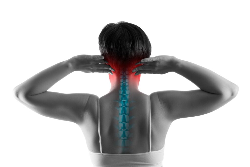
Occipital Headaches
Chiari headaches are felt at the occiput – at the base of the back of the skull and upper neck. They are generally tussive in nature, where they are exacerbated by valsalva maneuvers, which generally include: coughing, sneezing, heaving, laughing hard, or bearing down (like with a bowel movement or childbirth). These maneuvers reduce cardiac output (the amount of blood coming from the heart with each heartbeat), which in turn affects the attempted flow of cerebrospinal fluid, and it increases vagal stimuli.
Occipital headaches occur at the back of the lower skull (occiput) and upper neck, on one or both sides of the upper spinal cord.
For more on Chiari Malformation: http://chiaribridges.org//chiari-malformation/
For an expansive review of the name and definition of Chiari Malformation: http://chiaribridges.org//whats-in-a-name-chiari-malformation/
Connecting the Three Headaches
Untreated high pressure can cause cranial leaks (which leads to low pressure) – often accompanied by cerebrospinal fluid leaking through the nose or ears.
CSF leaks can sometimes seal on their own leading to rebound high pressure (which is temporary) or continued high pressure if they originally had high pressure.
Untreated high pressure can push the cerebellar tonsils down into the foramen magnum where it blocks the flow of cerebrospinal fluid, leading to occipital headaches (often diagnosed as a Chiari 1 Malformation) AND in return, the blockage of cerebrospinal fluid further increases intracranial pressure.
Untreated low pressure can cause the brain to sag and as it sags the cerebellar tonsils can get lodged into the foramen magnum where it blocks the flow of cerebrospinal fluid, leading to occipital headaches (often diagnosed as a Chiari 1 Malformation).
Untreated spinal leaks can create a suctioning or pulling down effect where the cerebellar tonsils can get lodged into the foramen magnum where it blocks the flow of cerebrospinal fluid, leading to occipital headaches (often diagnosed as a Chiari 1 Malformation).
All of the above is most common in patients with a connective tissue disorder such as Ehlers-Danlos Syndrome.
Major Problem Regarding Our Diagnoses & Treatment Options:
Doctors and radiologists alike, tend to see the herniated tonsils and assume a small posterior fossa.
Most do not check for high pressure or low pressure, even when directly asked and symptoms are present.
When a posterior fossa decompression is finally offered, the high or low pressure is often left untreated which leads to a failed decompression.
By the time sufferers get a name to go with their symptoms, we jump at the opportunity for relief.
The “Bobble-head Sensation” – When It Feels Like Your Neck Can No Longer Hold Up Your Head
While most of us experience this feeling either intermittently or continuously, it is generally related to structural instability issues:
Craniocervical Instability (CCI, also known as Syndrome of Occipitoatlantialaxial Hypermobility) involves vertical hypermobility (back and forth sliding) of the craniocervical junction (interface between the occipital bone and the 1st and 2nd vertebrae), where the neck is no longer properly supporting the cranium. This condition can be dangerous as it often involves brain stem compression that can lead to a vast array of symptoms of Dysautonomia (dysfunction of the Autonomic Nervous System – ANS).
For more on Craniocervical Instability and Other Related Disorders: http://chiaribridges.org/craniocervical-instability-related-disorders/
For a list of common CCI symptoms: http://chiaribridges.org//glossary/symptoms-of-craniocervical-instability/
For a list of common AAI (a condition commonly seen with CCI) symptoms: http://chiaribridges.org//glossary/symptoms-of-atlantoaxial-instability/
Subaxial Instability (SAI; also known as Cervical Instability) involves hypermobility of the C2/C3 to the C7 intervertebral discs. This condition (like most conditions involving the cervical spine) is a major cause of muscle spasms (in the neck and throughout the body at any point below the disc issues. When these neck spasms occur, they can cause the “Bobble-head sensation” where it feels like your neck can no longer hold up your head. This disc degeneration can lead to paralysis as discs compress the spinal cord.
Important Questions to Ask Your Neurosurgeons: http://chiaribridges.org/important-questions-for-your-neurosurgery-appointment/
Cerebrospinal fluid (CSF) is the clear, colorless liquid that surrounds the brain and spinal cord and is contained within a lining called the dura. The cerebrospinal fluid protects and cushions the brain and central nervous system. Among other functions, this fluid provides buoyancy to the brain, allowing it to float and weigh less, thus reducing the pressure at the base of the brain. A cerebrospinal fluid (CSF) leak occurs when there is a tear or hole in the dura that then allows this fluid to escape[1]. When leaks occur, the overall volume and pressure within the skull drops, and the cushioning and buoyancy effect is reduced, causing the brain to slump. In many cases, this leads to a condition known as intracranial hypotension and a vast range of symptoms.
The main symptom of a CSF leak is a headache that is worse when upright and improves when lying down horizontally. This is sometimes called a “positional” or “orthostatic” headache. However, not all positional headaches can be attributed to a CSF leak, and not all CSF leak headaches are positional. This is particularly the case in the chronic (vs acute) phase of CSF leaks, where the “positional” or “orthostatic” characteristic of symptoms may become more constant, lessen, or disappear entirely, including headache. Symptoms often worsen as the day goes on. Other leak symptoms can include, but are not limited to: nausea, vomiting, neck pain or stiffness, heaviness of head, pain between the shoulder blades, feeling of pressure within the head, changes in hearing (muffled or underwater sensation), tinnitus (ringing, buzzing, or pulsatile), feeling of liquid in the ears, sense of imbalance, sensitivity to light, sensitivity to sound, pain or numbness in the arms, changes in cognition (“brain fog,” memory loss, or loss of concentration), dizziness or vertigo, scalp sensitivity or tingling sensation within the scalp, visual changes (blurring, double vision, visual field defects), pain behind the eyes or when moving eyes, facial numbness or pain, sinus pressure, temporomandibular joint pain and stiffness, and subdural hematoma[2]. Cranial leak specific symptoms can vary even more and can include: fluid discharge from ears, nose (usually only one side) and to back of throat often reported as salty or metallic tasting, recurring or chronic meningitis, loss of sense of smell, change in hearing or ringing in the ears, and less frequently cognitive changes. Rare signs or complications of CSF leaks can include: quadriplegia, dementia (often mimicking Frontotemporal Lobe Dementia), Parkinsonism, other movement disorders, ataxia (unsteady gait), hypersomnolence, stupor, coma, stroke (hemorrhagic or ischemic), and even death.
CSF leaks are often very hard to locate, if ever. Approximately 50% of leaks cannot be found on imaging. Imaging and other tests used to attempt to find leaks are often read as “normal” even when there is a leak present. Other times, especially (but not always) in the case of chronic leaks, the positional symptoms either lessen or go away altogether, including the headache. Many who are leaking are not even aware that they are leaking. Leaks are often misdiagnosed as well[3]. Some of those common misdiagnoses are Postural Orthostatic Tachycardia Syndrome (POTS), migraines, sinus headaches, Meniere’s Disease, Chronic Fatigue Syndrome, Parkinson’s Disease (sometimes other neurodegenerative diseases), Fibromyalgia, Ehlers-Danlos Syndrome, Tarlov Cyst, Chiari Malformation, Cervical Spine Disease, cervicogenic headache, tension headache, and Sinusitis. To make diagnosis even more complex and elusive, CSF leaks can and do often occur along with any of these disorders and perhaps several simultaneously. A leak can cause an acquired Chiari malformation or coexist and complicate an existing congenital Chiari malformation[4]. Some patients have had unnecessary decompression surgeries when the underlying, sole cause was a leak all along.
Leaks can be caused by:
Medical procedures (also called iatrogenic leaks) for various diagnostic or therapeutic reasons such as lumbar punctures to collect fluid for analysis if meningitis is suspected, lumbar puncture for injection of contrast (myelography), spinal anesthesia, epidural injections, epidural steroid injections, prior skull base or spinal surgery, CSF shunt over-drainage, prior sinonasal surgery, and chiropractic or other spinal manipulations.
Traumatic injuries such as brachial plexus injuries falls, sports injuries, motor vehicle accidents, roller coaster rides, and other whiplash injuries.
Spontaneous leaks that occur with minimal or no clear cause. Sometimes spontaneous leaks may be associated with some sort of spinal pathologies such as calcified disk material or bone spurs. These leaks are usually ventral (or in front of the spinal cord).
There is growing evidence suggesting that a significant number of spontaneous CSF leaks occur as the result of a preexisting weakness in the dura[5]. Heritable Disorders of Connective Tissues (HDCT’s) such as Marfan Syndrome, Ehlers-Danlos Syndrome (both classical and hypermobility type), autosomal dominant Polycystic Kidney Disease, and other HDCT’s predispose patients to CSF leaks. One leak expert estimates that “slightly less than 100% of patients with spontaneous CSF leaks have an underlying connective tissue disorder.”[6] The dura is made out of connective tissue and patients with HDCT’s have thinner dura mater, that is more susceptible to tears and leaks. HDTC patients are more prone to spinal conditions such as perineural cysts, meningeal diverticula, and other HDCT defects such as aneurysms and dilatations. Oftentimes, a CSF leak is the first sign of an underlying HDCT.
Lumbar punctures (LP’s or spinal taps) should be avoided in patients with Chiari Malformation and/or in patients with HDCT’s[7]. There is a risk of causing a herniation of the cerebellar tonsils or making an existing herniation worse from the pull-down mechanism involved in lumbar punctures. Unfortunately, lumbar punctures are not always avoidable and sometimes very necessary, especially in cases to rule out life-threatening viral or bacterial conditions such as meningitis, subarachnoid hemorrhage, encephalitis, or syphilis. In these cases, measures can be taken to minimize LP risks such as using a certain needle type and size, limit the number of cc’s collected (by spontaneous drip ONLY), and, of course, always done under fluoroscopy by a competent physician[8]. Additionally, it is important to be aware that patients with HDCT’s are at greater risk for the dura to fail to heal following an LP. Patient’s should be aware of post-dural puncture headache (PDPH) symptoms and speak with their physicians if they suspect a leak following an LP.
The procedures and tests used to diagnose leaks will vary between patients and certain criteria are used to diagnose leaks[9]. Some of these tests and procedures might be: endoscopic exam and fluid collection and Beta-2-Trasnferrin testing (cranial leaks), Cisternography (including radioisotope cisternogram), Magnetic Resonance Imaging (MRI) including Magnetic Resonance (MR) myelography, Dynamic CT myelography, Digital Subtraction myelography, and Intrathecal saline infusion-enhanced myelography, a lumbar puncture to collect and test fluid and measure opening pressure. This imaging often includes both brain and spinal imaging. The normal opening pressure is not uncommon and does not rule out a leak. High pressure can also occur while leaking. Pre-existing intracranial hypertension can be related to the development of spontaneous spinal CSF leaks. Some reports suggest that spontaneous cerebrospinal fluid (CSF) leaks are strongly associated with idiopathic intracranial hypertension (IIH). There are 5 main findings on imaging that doctors look for, however, the absence of these findings does not rule out a CSF leak. The mnemonic SEEPS is used for most of these findings: subdural fluid collection, enhancement of pachymeninges, engorgement of venous structures, pituitary hyperemia, and sagging of the brain[10]. Other imaging findings that might be seen are small ventricles, cisterns might have less fluid, optic chasm might flatten over pituitary, pituitary might enlarge, empty sella, fluid in front of the pons, or pons might become flatter than normal. Repeat imaging is often necessary.
Treatment of leaks can either be medical or surgical. Conservative treatment is often recommended, if possible. This can include bed rest and avoidance of coughing, sneezing, straining, bending, twisting, and lifting, increased fluid intake and caffeine, the use of an abdominal binder, and sometimes steroids are recommended. Others may have a lumbar drain placed in the low back to decrease the pressure of the CSF around the area of the leak in an attempt to allow this area to heal. Some patients may need an epidural blood patch. Where a blood patch is not successful, a fibrin glue patch may be tried. About 30-40% of leaks occur at multiple sites, especially in those with an HDCT. Multisite patches may be required. Higher volumes of blood may be needed in order to reach where it needs to go, and/or the position of the needle may need to vary from the standard placement (transparietal or lateral placement)[11]. It is not uncommon for several patches to be tried. Many doctors make the mistake that if an EBP fails, there was no leak as well. Sometimes when more conservative and less invasive treatments have failed, neurosurgery may be necessary. Surgical repairs vary and are tailored according to the type and location of the leak. Sometimes in a select set of patients, other procedures have been used including epidural saline infusions through an implanted epidural catheter or lumbar dural reduction surgery. A condition known as rebound intracranial hypertension (RHP) may occur following any of these treatments[12]. Usually, but not always, there is a different pattern to the headache where one feels worse when horizontal and better when upright. Sometimes, acetazolamide (Diamox) or a similar medication is prescribed to help treat RHP.
Leaks are poorly recognized, poorly understood, under-researched, understudied, often misdiagnosed, can complicate existing conditions, are difficult to find, mimic many other disorders (including Chiari Malformation), and can be comprised of a vast array of symptoms. Most doctors are familiar with the symptoms of a leak in the acute phase. Very few doctors are familiar with how long-term, chronic CSF leaks “present” in regard to headaches and other leak symptoms and often miss the more subtle symptoms of chronic leaks. Like Chiari and other related disorders, no two patients with CSF leaks have the same symptoms and often experience misdiagnosis, delayed diagnosis, are disbelieved concerning their symptoms of the severity thereof, and all too often dismissed to suffer excruciating pain, decline, and debility. Educating yourself as much as possible about CSF leaks will help guide and empower you and those around you who may have existing, suspected or potential future complications that may arise due to CSF leaks.
5 Reinstein, Eyal, et al. “Connective Tissue Spectrum Abnormalities Associated with Spontaneous Cerebrospinal Fluid Leaks: a Prospective Study.” European Journal of Human Genetics, Nature Publishing Group, Apr. 2013, <www.ncbi.nlm.nih.gov/pmc/articles/PMC3598315/>.
7 Erbay, Sami H., et al. “Is Lumbar Puncture Contraindicated in Patients with Chiari I Malformation?” American Journal of Neuroradiology, American Journal of Neuroradiology, 1 Apr. 2005, <www.ajnr.org/content/26/4/985>.
9 Schievink, W. l., et al. “Diagnostic Criteria for Spontaneous Spinal CSF Leaks and Intracranial Hypotension.” American Journal of Neuroradiology, American Journal of Neuroradiology, 1 May 2008, <www.ajnr.org/content/29/5/853>.
11 Griauzde, J., et al. “Large-Volume Blood Patch to Multiple Sites in the Epidural Space through a Single-Catheter Access Site for Treatment of Spontaneous Intracranial Hypotension.”American Journal of Neuroradiology, American Journal of Neuroradiology, 30 Apr. 2014, <www.ajnr.org/content/early/2014/04/30/ajnr.A3945>.
12 Kranz, P. G., et al. “Rebound Intracranial Hypertension: A Complication of Epidural Blood Patching for Intracranial Hypotension.” American Journal of Neuroradiology, American Journal of Neuroradiology, 1 June 2014, <www.ajnr.org/content/35/6/1237>.